Coupled hair cells in the inner ear – „Together we are strong!“
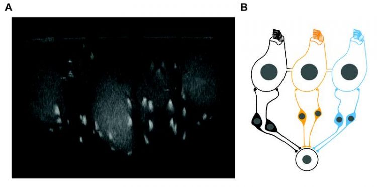
Figure: Coupled hair cells Source: Jean et al., Nat Commun, 2020; Suppl. Part
Hearing is one of our most important senses. Hearing disorders are very common: according to the World Health Organization (WHO), about 466 million people (five percent of the world's population) suffer from hearing loss and require medical treatment. It is therefore of utmost important to understand the basic processes of hearing
Until now, it has assumed that the sensory cells of the cochlea, the so called inner hair cells, are electrically strictly separated from each other, thus achieving maximum pitch or frequency resolution.
A team of scientists from the Institute for Auditory Neuroscience and the Department of Otorhinolaryngology at the University Medical Center Göttingen (UMG) as well as the Max Planck Institute of Experimental Medicine in Göttingen, the Göttingen Campus Institute for Dynamics of Biological Networks and the University College London have now shown in a multidisciplinary approach that this assumption is not always correct in the cochlea of adult rodents.
Instead, 30 percent of the cells form an electrical and chemical unit with an average of three inner hair cells that jointly get excited to transmit the acoustic signal. These research findings were obtained within the framework of the Collaborative Research Center 889 “Cellular Mechanisms of Sensory Processing” and the Cluster of Excellence “Multiscale Bioimaging”, and published in the renowned journal “Nature Communications”.
Original publication: Jean P, Anttonen T, Michanski S, de Diego A, Steyer AM, Neef A, Oestreicher D, Kroll J, Nardis C, Pangršič T, Möbius W, Ashmore J,
Wichmann C, Moser T (2020) Macromolecular and electrical coupling between inner hair cells in the rodent cochlea. Nat Commun, 11, 3208 (2020). DOI: https://doi.org/10.1038/s41467-020-17003-z or: https://rdcu.be/b5aGS
Detailed research results
In adult animals, about one third of the hair cells are part of mini-electrochemically connected in “mini-syncytia”, which have been identified for the first time, and are distributed throughout the cochlea.
Using high-resolution fluorescence microscopy, the scientists showed that the ribbon synapses of all cells in such mini-syncytia react together upon stimulation of one single cell. The coupling of hair cells allowed dyes and metabolic products to distribute freely within these cell units (see figure).
However, this electrical and chemical coupling is not mediated via the widely-used protein pores between neighbouring cells, the so-called gap junctions. Apparently, the hair cells employ an unconventional mechanism of cell coupling: Innovative 3-dimensional electron microscopy indicated that the cell envelopes of neighbouring cells fuse.
The scientists were able to show that even very large molecules, such as antibodies, or small cell organelles can pass between the cells. “This study opens an exciting new perspective in sensory research”, says Prof. Dr. Carolin Wichmann, head of a research group at the Institute for Auditory Neurosciences, UMG, and project leader in the Collaborative Research Center 889 and the Cluster of Excellence “Multiscale Bioimaging”.
“This phenomenon needs further investigation in order to clarify the molecular mechanism of coupling, its importance for the sense of hearing and its occurrence in the inner ear of other animal species,” says Wichmann.
Computer simulations also suggest that the processing of weak sound signals is improved by the formation of hair cell mini-syncytia. “The organisation in mini-syncytia makes the sensory function more sensitive and at the same time more robust.
Thus, even if a hair cell is damaged in its sensitive hair bundle, it can be 'dragged along' by its neighbouring cells and thus contribute to sound processing”, says first author Dr. Philippe Jean, who received his doctorate in the sensory research doctoral program of the Göttingen Graduate Center for Neurosciences, Biophysics and Molecular Biosciences (GGNB) and is now researching at the Institut de l'Audition in Paris.
“The pitch resolution of our hearing is apparently not reduced by the limited number of cells in the cell assemblies”, adds Jean.
“This study is a very good example of the possibilities that multiscale imaging opens up in the new Göttingen Cluster of Excellence”, says Prof. Dr. Tobias Moser, Director of the Institute for Auditory Neuroscience, UMG, and spokesperson of the “Multiscale Bioimaging” Cluster of Excellence and the Collaborative Research Center 889 “Cellular Mechanisms of Sensory Processing”.
“Only by combining light and electron microscopy these processes could be studied over scales of a few billionths of a meter to about 100 millionths of a meter, and the probable coupling mechanism could be unraveled”.
Background Information about hearing
Hearing begins with the sound being captured in the auricle, the acoustic wave is transmitted, causing the eardrum and ossicles in the middle ear to move to convey the signal to the inner ear. Here, the inner hair cells are located and convert the sound waves into signals, which can be read by the brain.
The inner hair cells are lined up like steps on a spiral staircase, with each step being assigned to a certain pitch level. The sensory signal is generated at the hair bundles of the inner hair cells: The mechanical opening of small protein pores (ion channels) leads to elec-trical recharging of the cell.
This apparatus is so sensitive that we can hear mechanical deflection of the hairs by the diameter of a hydrogen atom. In the basal part of inner hair cells the communication units between hair cells and the nerve fibers, the so-called ribbon synapses, are located. Ribbon synapses are, nanometer-sized structures that enable a sustained release of messenger from synaptic vesicles.
Further Information:
Institute for Auditory Neuroscience:
http://www.auditory-neuroscience.uni-goettingen.de/
Collaborative Research Center 889 “Cellular Mechanisms of Sensory Processing”: www.sfb889.uni-goettingen.de
Cluster of Excellence “Multiscale Bioimaging”: https://mbexc.de/
The Göttingen Cluster of Excellence “Multiscale Bioimaging: From Molecular
Machines to Networks of Excitable cells” (MBExC) is funded since January 2019 in the framework of the Excellence Strategy of the German Federal and State Government. Applying a unique and multiscale approach, MBExC investigates the disease-relevant functional units of electrically active cells of heart and brain, from the molecular to the organ level. The MBExC unites numerous partners from the university and extra-university institutions in Göttingen. The overall goal: to under-stand the relationship between heart and brain diseases, to link basic and clinical research, and thus to develop new therapeutic and diagnostic approaches with social implications.
https://mbexc.de/
Figure: Coupled hair cells (left): Fluorescence microscopy image of a cell cluster of five coupled inner hair cells (pear-shaped with bright spots). The second cell from the left was loaded with a fluorescent dye that labels the synaptic ribbon using the patch-clamp technique. From there, the dye spreads into the neighbouring hair cells and marks the synaptic ribbons (bright spots). Source: Institute for Auditory Neuroscience / UMG. (right) A “mini-syncytium” of three coupled inner hair cells (the connection is simplified as a tube formed by fused cell membranes). The electrical signals are transmitted via coupling of the cells. This mechanism reduces the stochastic noise of signal transmission at the ribbon synapses with the neurons forming the auditory nerve. From there the information is transmitted to a common target neuron in the brain, further reducing the noise and resulting in higher hearing sensitivity.
Source: Jean et al., Nat Commun, 2020; Suppl. Part.
Contact
University Medical Center Göttingen, University of Göttingen
Institute for Auditory Neuroscience
Prof. Dr. Tobias Moser
Robert-Koch-Str. 40, 37075 Göttingen
phone 0551 / 39-63071
tmoser@gwdg.de
Institute for Auditory Neuroscience
Center for Biostructural Imaging of Neurodegeneration
Prof. Dr. Carolin Wichmann
Von-Siebold-Str. 3a, 37075 Göttingen
phone 0551 / 39-61128
carolin.wichmann@med.uni-goettingen.de
Original publication: Jean P, Anttonen T, Michanski S, de Diego A, Steyer AM, Neef A, Oestreicher D, Kroll J, Nardis C, Pangršič T, Möbius W, Ashmore J,
Wichmann C, Moser T (2020) Macromolecular and electrical coupling between inner hair cells in the rodent cochlea. Nat Commun, 11, 3208 (2020). DOI: https://doi.org/10.1038/s41467-020-17003-z or: https://rdcu.be/b5aGS
Media Contact
More Information:
https://www.umg.euAll latest news from the category: Health and Medicine
This subject area encompasses research and studies in the field of human medicine.
Among the wide-ranging list of topics covered here are anesthesiology, anatomy, surgery, human genetics, hygiene and environmental medicine, internal medicine, neurology, pharmacology, physiology, urology and dental medicine.


Newest articles

Silicon Carbide Innovation Alliance to drive industrial-scale semiconductor work
Known for its ability to withstand extreme environments and high voltages, silicon carbide (SiC) is a semiconducting material made up of silicon and carbon atoms arranged into crystals that is…

New SPECT/CT technique shows impressive biomarker identification
…offers increased access for prostate cancer patients. A novel SPECT/CT acquisition method can accurately detect radiopharmaceutical biodistribution in a convenient manner for prostate cancer patients, opening the door for more…

How 3D printers can give robots a soft touch
Soft skin coverings and touch sensors have emerged as a promising feature for robots that are both safer and more intuitive for human interaction, but they are expensive and difficult…

