Cellular mechanisms of early mammary gland development unraveled
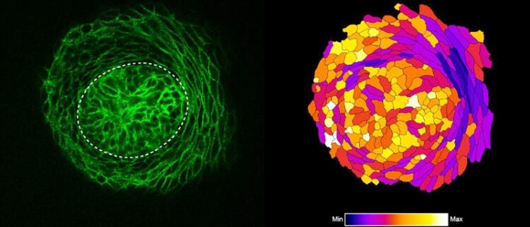
Optical section of a confocal microscopy image of the mammary bud (marked with dashed line) stained with an epithelial marker in green at the time when ring cells form (left) and analysis of cell roundness of segmented cells with ring cells in dark blue (right).
Credit: Ewelina Trela
Helsinki University research group used live tissue imaging for the first time to visualise the emergence of the mammary gland.
Despite long-standing interest, the cellular mechanisms driving the initiation of mammary gland development have remained elusive for decades, mostly due to technical limitations in studying dynamic cell behaviors in live tissues. Recent advances in microscopic methods and availability of various mouse models allowed the research group of Marja Mikkola from HiLIFE Institute of Biotechnology, University of Helsinki to address this question. This is the first time when live tissue imaging has been used to visualise the emergence of the mammary gland.
Mammary gland is the class-defining organ of mammals, yet we know surprisingly little how its development commences. In their recent study published in Journal of Cell Biology, the research group of Marja Mikkola used time-lapse imaging to show that the growth of the mammary bud is primarily fueled by migration of cells to the bud. In contrast, although increase in cellular size and cell proliferation contribute to this process, the role of these mechanisms remains minor.
“Interestingly, mammary bud cells, unlike most of other skin derivatives such as hair follicle and tooth bud, do not divide for several days, indicating that this might be a unique feature of early mammary gland development” says graduate student Ewelina Trela, the lead author of the study. “However, we do not yet know why this happens”, she continues.
Mammary buds use a previously undescribed mechanism for invagination
Tissue invagination, or tissue folding inwards into the underlying stroma, is a fundamental mechanism that occurs to generate the architecture of many organs. In the same piece of work, the authors describe a novel mechanism for tissue invagination.
“Using confocal fluorescence microscopy, we found thin and elongated epidermal keratinocytes surrounding mammary bud in a rim like fashion: their appearance and disappearance coincided with the invagination process suggesting that these cells, named ring cells, could be functionally important” details principal investigator Marja Mikkola.
Next, the Mikkola group teamed up with the group of Sara Wickström at HiLIFE and Faculty of Medicine, University of Helsinki to establish live imaging of the forming mammary bud, which confirmed that ring cells move circumferentially around the mammary bud.
The study also revealed that the ring cells exert contractile force through the actomyosin network, via non-muscle myosin IIA (NMIIA). The functionality of ring cells was impaired in NMIIA deficient mice leading to compromised mammary bud shape. Whether other developing organs utilize a similar cellular mechanism for invagination remains an open question.
All latest news from the category: Life Sciences and Chemistry
Articles and reports from the Life Sciences and chemistry area deal with applied and basic research into modern biology, chemistry and human medicine.
Valuable information can be found on a range of life sciences fields including bacteriology, biochemistry, bionics, bioinformatics, biophysics, biotechnology, genetics, geobotany, human biology, marine biology, microbiology, molecular biology, cellular biology, zoology, bioinorganic chemistry, microchemistry and environmental chemistry.


Newest articles

Silicon Carbide Innovation Alliance to drive industrial-scale semiconductor work
Known for its ability to withstand extreme environments and high voltages, silicon carbide (SiC) is a semiconducting material made up of silicon and carbon atoms arranged into crystals that is…

New SPECT/CT technique shows impressive biomarker identification
…offers increased access for prostate cancer patients. A novel SPECT/CT acquisition method can accurately detect radiopharmaceutical biodistribution in a convenient manner for prostate cancer patients, opening the door for more…

How 3D printers can give robots a soft touch
Soft skin coverings and touch sensors have emerged as a promising feature for robots that are both safer and more intuitive for human interaction, but they are expensive and difficult…

