Cell biology: Signal transduction without signal
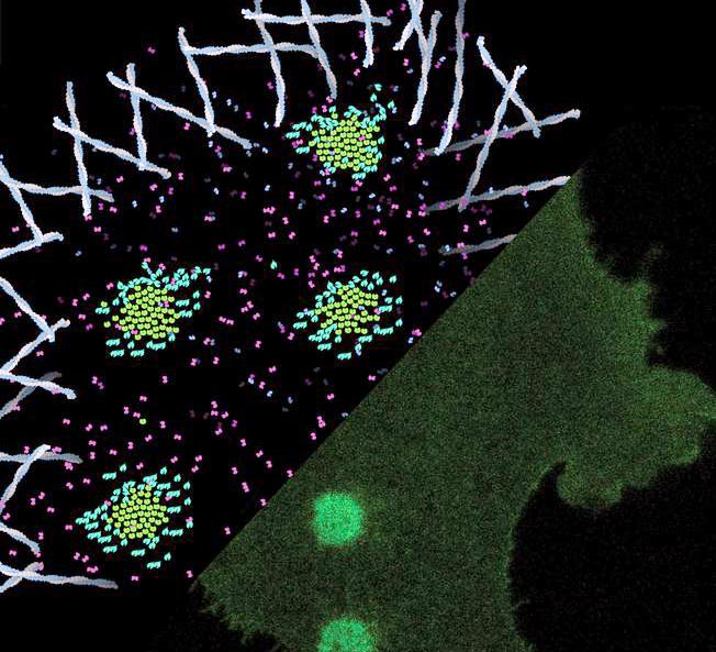
Laser spots activate very small synthetic lock-and-key pairs in a matrix to create receptor clusters in the cell membrane. This ligand-independent activation triggers calcium signaling and increased cell motility.
M. Florencia Sánchez & Robert Tampé, Goethe University Frankfurt
– receptor clusters can direct cell movement
Whether we smell, taste or see, or when adrenaline rushes through our veins, all of these signals are received by our cells via a specific group of receptor proteins called G protein-coupled receptors, which transmit signals to the inside of the cell. Biochemists at Goethe University Frankfurt and the University of Leipzig have now discovered that such receptors can also produce signals even in the absence of an external stimulus: It is apparently sufficient for certain receptors if many of them are clustered at the cell surface. (Science, doi/10.1126/science.abb7657)
Our body consists of 100 trillion cells that communicate with each other, receive signals from the outside world and react to them. A central role in this communication network is attributed to receiver proteins, called receptors, which are anchored at the cell membrane. There, they receive and transmit signals to the inside of the cell, where a cell reaction is triggered.
In humans, G protein-coupled receptors (GPC receptors) represent the largest group of these receptor molecules, with around 700 different types. The research of the Frankfurt and Leipzig scientists focused on a GPC receptor that serves as a receptor for the neuropeptide Y in cells and is accordingly called the Y2 receptor. Neuropeptide Y is a messenger substance that primarily mediates signals between nerve cells, which is why Y2 receptors are mainly present in nerve cells and among other activities trigger the formation of new cell connections.
In the laboratory, the researchers engineered cells, which had approx. 300,000 Y2 receptors on their surface and were grown on specifically developed, light-sensitive matrices. Each of the Y2 receptors was provided with a small molecular “label”. Once the scientists created a spot of light with a fine laser beam on the cell surface, the Y2 receptor under this spot were trapped via the molecular label to the exposed matrix in such a way that the Y2 receptors moved closely together to form an assembly known as a cluster. The whole reaction could be immediately observed at the defined spot and within a few seconds.
Professor Robert Tampé from the Institute of Biochemistry at Goethe University Frankfurt explains: “The serendipity about this experiment is that the clustering of receptors triggers a signal that is similar to that of neuropeptide Y. Solely by the clustering, we were able to trigger cell movement as a reaction of the cell. The laser spots even allowed us to control the direction of the cell movement.” As the light-sensitive lock-and-key pairs utilized are very small compared to the receptors, the organization of the receptors in the cell membrane can be controlled with high precision using the laser spot. “This non-invasive method is thus particularly well suited to study the effects of receptor clustering in living cells,” Tampé continues. “Our method can be used to investigate exciting scientific questions, such as how receptors are organized in networks and how new circuits are formed in the brain.”
Image/Movie downloads:
http://www.uni-frankfurt.de/98160408
Caption Image: Laser spots activate very small synthetic lock-and-key pairs in a matrix to create receptor clusters in the cell membrane. This ligand-independent activation triggers calcium signaling and increased cell motility. (Graphic copyright: M. Florencia Sánchez & Robert Tampé, Goethe University Frankfurt.)
http://www.uni-frankfurt.de/98150564
Caption Movie: Upon irradiation with laser light (white rings), receptors cluster in the cell (light green circles). Thereupon, the cell moves into the direction of the receptor clusters. (Copyright: M. Florencia Sánchez & Robert Tampé, Goethe University Frankfurt). Reprinted with permission from M. F. Sánchez et al., Science 10.1126/science.abb7657(2021).
Wissenschaftliche Ansprechpartner:
Professor Robert Tampé
Institute of Biochemistry
Goethe-Universität Frankfurt, Germany
Phone: +49 69 798 29475
tampe@em.uni-frankfurt.de
http://www.biochem.uni-frankfurt.de/
Originalpublikation:
M. Florencia Sánchez, Sylvia Els-Heindl, Annette G. Beck-Sickinger, Ralph Wieneke, Robert Tampé: Photo-induced receptor confinement drives ligand-independent GPCR signaling. Science abb7657
DOI: 10.1126/science.abb7657; https://science.sciencemag.org/lookup/doi/10.1126/science.abb7657
Media Contact
All latest news from the category: Life Sciences and Chemistry
Articles and reports from the Life Sciences and chemistry area deal with applied and basic research into modern biology, chemistry and human medicine.
Valuable information can be found on a range of life sciences fields including bacteriology, biochemistry, bionics, bioinformatics, biophysics, biotechnology, genetics, geobotany, human biology, marine biology, microbiology, molecular biology, cellular biology, zoology, bioinorganic chemistry, microchemistry and environmental chemistry.


Newest articles

High-energy-density aqueous battery based on halogen multi-electron transfer
Traditional non-aqueous lithium-ion batteries have a high energy density, but their safety is compromised due to the flammable organic electrolytes they utilize. Aqueous batteries use water as the solvent for…

First-ever combined heart pump and pig kidney transplant
…gives new hope to patient with terminal illness. Surgeons at NYU Langone Health performed the first-ever combined mechanical heart pump and gene-edited pig kidney transplant surgery in a 54-year-old woman…

Biophysics: Testing how well biomarkers work
LMU researchers have developed a method to determine how reliably target proteins can be labeled using super-resolution fluorescence microscopy. Modern microscopy techniques make it possible to examine the inner workings…

