X-rays in a new light

Visible light and X-rays are different types of radiation. Visible light, for example, doesn’t penetrate the human body, whereas X-rays are absorbed weakly and can be used in medical imaging.
Similar differences exist at very high light intensities, which make X-rays potentially useful in materials science, but this area—referred to as ‘nonlinear optics’—remains largely unexplored. Now, researchers from the RIKEN SPring-8 Center in Harima have taken the first step in establishing a more systematic approach to studying nonlinear X-ray effects1.
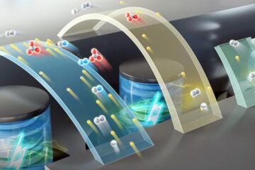
The team investigated the so-called parametric down-conversion of a single X-ray photon that splits into two separate photons, whose combined energy equals the original photon’s energy. This effect was studied in a diamond crystal, which provided the medium for this process to occur. The necessary high intensity X-ray radiation came from the SPring-8 synchrotron, which is ideally suited for the task, according to Kenji Tamasaku from the research team. “It delivers some of the world’s brightest X-rays.”
However, a competing process can occur in addition to the down-conversion: the creation of only one X-ray photon and the simultaneous excitation of one of the material’s electron to another state from the remainder of the original energy. An observer cannot distinguish which of these processes actually occurred in the material to produce outcoming photons of the same energy, which means that there is a quantum mechanical interference between both processes. This is known as the Fano effect.
Tamasaku and colleagues studied the Fano effect for a range of parameters including X-ray photon energy. Based on theoretical modeling of a large dataset available from their experiments, they quantified efficiency of the nonlinear down-conversion process of X-rays for the first time. The possibility of this achievement had long been doubtful, as it requires not only a careful experimental calibration, but also very high X-ray intensities that are available at SPring-8.
The quantitative results for the nonlinear optical parameters of the down-conversion process are convincing and largely in line with theoretical expectations, even though some of the features observed remain poorly understood.
Nevertheless, Tamasaku is confident that “these results represent the first firm base from which to venture into the frontier of X-ray nonlinear optics.” In particular, he is hopeful that the completion of a new X-ray laser called X-ray Free Electron Laser (XFEL) at SPring-8 next year will significantly expand the potential for the study of these non-linear optical effects.
The corresponding author for this highlight is based at the Coherent X-Ray Optics Laboratory, RIKEN SPring-8 Center
Media Contact
All latest news from the category: Physics and Astronomy
This area deals with the fundamental laws and building blocks of nature and how they interact, the properties and the behavior of matter, and research into space and time and their structures.
innovations-report provides in-depth reports and articles on subjects such as astrophysics, laser technologies, nuclear, quantum, particle and solid-state physics, nanotechnologies, planetary research and findings (Mars, Venus) and developments related to the Hubble Telescope.
Newest articles

High-energy-density aqueous battery based on halogen multi-electron transfer
Traditional non-aqueous lithium-ion batteries have a high energy density, but their safety is compromised due to the flammable organic electrolytes they utilize. Aqueous batteries use water as the solvent for…

First-ever combined heart pump and pig kidney transplant
…gives new hope to patient with terminal illness. Surgeons at NYU Langone Health performed the first-ever combined mechanical heart pump and gene-edited pig kidney transplant surgery in a 54-year-old woman…

Biophysics: Testing how well biomarkers work
LMU researchers have developed a method to determine how reliably target proteins can be labeled using super-resolution fluorescence microscopy. Modern microscopy techniques make it possible to examine the inner workings…


