What lies beneath: mapping hidden nanostructures
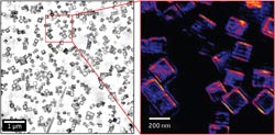
Figure 1: Images of gold/silver nanoparticles, acquired using a combined method of x-ray ptychography and anomalous x-ray diffraction. Copyright : 2012 Yukio Takahashi<br>
The ability to diagnose and predict the properties of materials is vital, particularly in the expanding field of nanotechnology. Electron and atom-probe microscopy can categorize atoms in thin sheets of material, and in small areas of thicker samples, but it has proven far more difficult to map the constituents of nanostructures inside large, thick objects. X-rays—the most common imaging tool for hard biological materials such as bones—have a limited focal-spot size, so they cannot focus on nanoscale objects.
Now, Yukio Takahashi and colleagues at Osaka University, together with researchers at Nagoya University and the RIKEN SPring-8 center in Hyogo, have succeeded for the first time in producing two-dimensional images of nanostructures encased in thick materials on a large scale1. Their work was possible because they designed a new x-ray diffraction microscopy system that does not require a lens.
“The main challenges in this work were to realize x-ray diffraction microscopy with a high resolution and a large field of view, then extend it to element-specific imaging,” Takahashi explains. “We achieved this by establishing a scanning x-ray diffraction imaging technique called x-ray ptychography.”
Ptychography involves taking images of an object that overlap with one another on a series of coinciding lattice points. The researchers combined this technique with x-rays, and included a system to compensate for the drifting of optics during imaging. Takahashi and his colleagues focused the x-rays using so-called ‘Kirkpatrick–Baez mirrors’ that allowed them to collect high-quality diffraction data.
Their system monitors the changes in the diffraction of x-rays at two different energies. The degree of phase difference between the two x-ray energies changes significantly at the absorption edge of the target element. This is related to the atomic number of the element, meaning that the elements present in the material can be identified. To verify that their system works, the researchers deposited gold/silver nanoparticles around 200 nanometers in size on a silicon nitride membrane, and produced high-resolution and large-scale images of the particles. The resolutions were better than 10 nanometers (Fig. 1).
“One of the practical applications [of this technique] in future is the possible observation of cells,” explains Takahashi. “The shape of a whole cell and the spatial distribution of [its] organelles could be three-dimensionally visualized at 10 nanometer resolution—to provide key insights into the organization inside cells. We hope to see this technique being used in biological and materials science in future.”
The corresponding author for this highlight is based at the SR Imaging Instrumentation Unit, RIKEN SPring-8 Center
Media Contact
All latest news from the category: Physics and Astronomy
This area deals with the fundamental laws and building blocks of nature and how they interact, the properties and the behavior of matter, and research into space and time and their structures.
innovations-report provides in-depth reports and articles on subjects such as astrophysics, laser technologies, nuclear, quantum, particle and solid-state physics, nanotechnologies, planetary research and findings (Mars, Venus) and developments related to the Hubble Telescope.


Newest articles

Silicon Carbide Innovation Alliance to drive industrial-scale semiconductor work
Known for its ability to withstand extreme environments and high voltages, silicon carbide (SiC) is a semiconducting material made up of silicon and carbon atoms arranged into crystals that is…

New SPECT/CT technique shows impressive biomarker identification
…offers increased access for prostate cancer patients. A novel SPECT/CT acquisition method can accurately detect radiopharmaceutical biodistribution in a convenient manner for prostate cancer patients, opening the door for more…

How 3D printers can give robots a soft touch
Soft skin coverings and touch sensors have emerged as a promising feature for robots that are both safer and more intuitive for human interaction, but they are expensive and difficult…

