Fixing of the lumbar column aided by simple radiological techniques
Fixing of the lumbar vertebral column aided by simple radiological techniques facilitates the process and avoids complications. This technique has arisen as a result of the conclusion of the PhD thesis by Dr. Matías Alfonso, specialist in the Department of Orthopaedic and Bone Surgery at the University Hospital of Navarre, and has been based on research carried out on a pedicular screw method based on intraoperatorial anatomical references. The study has been applied to 44 patients attending the University Hospital.
The insertion of pedicular screws into the lumbar vertebral column is suitable for the treatment of fractures, problems of instability due to degenerative illness, tumours, etc. It involves a routine technique in orthopaedics and bone surgery. Nevertheless, lesion of the nerve roots can occur as a complication, a risk that can affect up to 3% of patients.
The University Hospital has evaluated a method to guarantee the pedicular screw system in a reliable way, avoiding the neurological complications. This system groups together a series of radiological projections by means of which we can see to the borders of the vertebrae, thus enabling the path of the screws to be ascertained with precision.
Adventages
The PhD research undertaken by Dr. Alfonso confirms that the pedicular screw system based on the radiological method obtains results similar to those produced by computer-assisted surgery. Moreover, the added advantage is that it is a much simpler procedure that has no need of neuronavigators, thus considerably reducing health costs.
Another of the study’s conclusions related to the shape of the vertebrae and the location of the injury. The method used in order to evaluate, in a preoperational manner, the shape and size of the vertebrae, enable us to know the width and length of the screws that we have to use and in which direction they have to go.
Finally, this research has demonstrated its usefulness in reliably determining the position of the screws once introduced and terminated the operation. After analysing the appearance of the inside of the vertebra, we have observed that 5.5 mm width screws appear as measuring up to 8 mm on the scanner. That is to say, our method has detected that the TAC image obtained can show that part of the screw is outside the vertebra when, in reality, this is not the case!
Media Contact
More Information:
http://www.basqueresearch.comAll latest news from the category: Health and Medicine
This subject area encompasses research and studies in the field of human medicine.
Among the wide-ranging list of topics covered here are anesthesiology, anatomy, surgery, human genetics, hygiene and environmental medicine, internal medicine, neurology, pharmacology, physiology, urology and dental medicine.


Newest articles
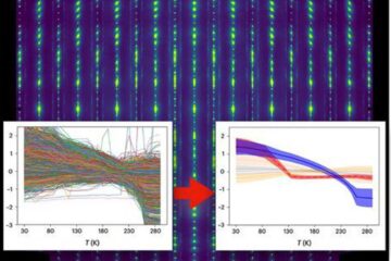
Machine learning algorithm reveals long-theorized glass phase in crystal
Scientists have found evidence of an elusive, glassy phase of matter that emerges when a crystal’s perfect internal pattern is disrupted. X-ray technology and machine learning converge to shed light…
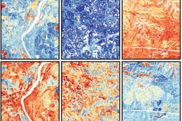
Mapping plant functional diversity from space
HKU ecologists revolutionize ecosystem monitoring with novel field-satellite integration. An international team of researchers, led by Professor Jin WU from the School of Biological Sciences at The University of Hong…
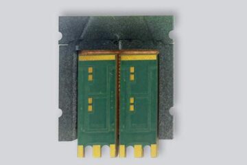
Inverters with constant full load capability
…enable an increase in the performance of electric drives. Overheating components significantly limit the performance of drivetrains in electric vehicles. Inverters in particular are subject to a high thermal load,…

