3D MDCT can replace conventional angiography of extremities
Three-dimensional MDCT angiography can be used in place of conventional angiography to image the extremities in nearly any case where conventional angiography is indicated, a new study suggests.
The study included 40 patients who underwent extremity MDCT angiography for a wide range of diseases, including traumatic injuries, musculoskeletal masses, and atherosclerotic disease, said lead author Dr. Musturay Karcaaltincaba of Hacettepe University School of Medicine in Ankara, Turkey. Diagnostic images were obtained in all patients, said Dr. Karcaaltincaba.
“There are a number of advantages of MDCT angiography – the study can be completed within minutes; it does not require sedation and arterial catheterization. Patients can go home right after the procedure. In addition, newer MDCT systems require a lower dose of contrast media, said Dr. Karcaaltincaba.
MDCT angiography can show fractured bones as well as vascular injury, which can reduce the number of X ray examinations the patient needs, thus speeding up the diagnosis. This is particularly important in the emergency room.
Musculoskeletal masses and the vasculature near the masses can be easily identified. This is important before biopsy or surgery to avoid hemorrhagic complications, he said. “3D images of the relevant anatomy (both vascular and musculoskeletal) provides surgeons with vivid images very similar to what they see during surgery and facilitates preoperative planning,” Dr. Karcaaltincaba said.
“Recent comparative studies emphasize the diagnostic power of this technique as an alternative to conventional angiography in diagnosing atherosclerosis,” he said. “Calcific plaques can be clearly seen,” he added.
There are some patients who cannot undergo an MDCT angiography examination – those with poor renal function or those who have a history of reaction to contrast material, Dr. Karcaaltincaba said. “Image quality is suboptimal in patients with metallic implants,” he added.
“MDCT angiography is currently the method of choice at our institution for imaging the arteries in the extremities. Although conventional angiography is still the gold standard, it is invasive, and we generally reserve it for interventional procedures,” he said. The study of appears in the July, 2004 issue of the American Journal of Roentgenology.
Media Contact
More Information:
http://www.arrs.orgAll latest news from the category: Health and Medicine
This subject area encompasses research and studies in the field of human medicine.
Among the wide-ranging list of topics covered here are anesthesiology, anatomy, surgery, human genetics, hygiene and environmental medicine, internal medicine, neurology, pharmacology, physiology, urology and dental medicine.


Newest articles
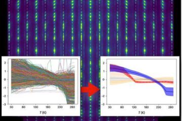
Machine learning algorithm reveals long-theorized glass phase in crystal
Scientists have found evidence of an elusive, glassy phase of matter that emerges when a crystal’s perfect internal pattern is disrupted. X-ray technology and machine learning converge to shed light…
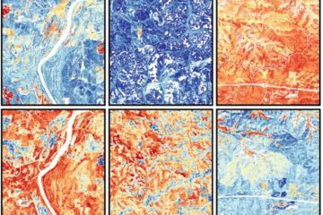
Mapping plant functional diversity from space
HKU ecologists revolutionize ecosystem monitoring with novel field-satellite integration. An international team of researchers, led by Professor Jin WU from the School of Biological Sciences at The University of Hong…

Inverters with constant full load capability
…enable an increase in the performance of electric drives. Overheating components significantly limit the performance of drivetrains in electric vehicles. Inverters in particular are subject to a high thermal load,…

