Three dimensional ecography
Three-dimensional ecography is a novel advance in the application of ultrasounds in the diagnosis of a number of pathologies. It involves a system of probes that register images in multiple layers. Then the information is transferred to a computer within the ecograph itself, where the three-dimensional reconstruction is carried out automatically. Advances in information technology have so perfected the data processing that this reconstruction can be obtained in real time.
This new diagnostic technique has an average duration of 15 minutes, although actually obtaining the images only takes a few seconds and has a number of advantages over the conventional bidimensional ecography, such as studying the possibility of studying all layers of the space, showing images in 3-D and obtaining data which is more reproducible.
Application in obstetrics
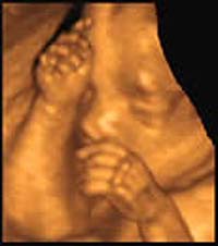
Three-dimensional ecography is considered a diagnostic technology a complementary to the conventional or bidimensional one which turns out to be highly useful for foetal study. It provides high-quality three-dimensional images of the foetus, to such an extent that parents can follow with precision the features and movements of their child in different positions and from different perspectives. According to some specialists, this enhancement of the image may bring greater bonding between parents and child.
Another application of 3-D ecography in the field of obstetrics is the detection of various foetal anomalies. More specifically, it provides additional information for the diagnosis of lesions of the face, the limbs and the vertebral column, and its clinical use in cardiac problems is being studied. This technique does not diagnose more anomalies than conventional ecography but rather establishes the degree of lesion with more certainty in order that the specialist can make an early prognosis of the most suitable form of therapy.
The University Hospital School has the most advanced equipment available on the market today, providing three-dimensional images of the foetus inside the uterus and with its movements. What we have here is 4th dimensional ecography, the main contribution of which being the study of intrauterine foetal behaviour. With this technique foetal movements will be able to be seen and foetal reactions to different stimuli, without using indirect methods such as the checking of foetal cardiac frequency.
Application in gynaecology
The studies carried out in the Department of Gynaecology and Obstetrics at the University Hospital School have confirmed that, with this technique, uterine malformations are diagnosed with more precision than with traditional ecography. Specifically, it modifies the diagnosis in 50% of the cases. i.e. it detects lesions that have not been diagnosed with conventional ecography and can also change the diagnosis from one anomaly to another. These lesions can give rise to repetitive abortions, difficulty in becoming pregnant or low birth weight births, conditions for which a proper diagnosis is fundamental in order to indicate appropriate treatment and in time. Another gynaecological anomaly which benefits from this imaging technique is with submucous miomas, treatment for which requires hysteroscopic resection. Three-dimensional ecography defines with more exactitude which cases are more suitable for treatment and guarantees post-operational results.
Apart from benign lesions, researchers at the University Hospital School are particularly interested in the value of three-dimensional ecography to the study of tumorous neoangiogenesis. Our preliminary data show that, with this technique, the degree of vascularisation of cancer of the neck of the uterus and of the endometrial tissue as well as their correlation with prognostic factors for the tumour, can be observed in an objective manner. It, in effect, involves information that can help to modify the treatment.
Media Contact
More Information:
http://www.unav.es/cunAll latest news from the category: Health and Medicine
This subject area encompasses research and studies in the field of human medicine.
Among the wide-ranging list of topics covered here are anesthesiology, anatomy, surgery, human genetics, hygiene and environmental medicine, internal medicine, neurology, pharmacology, physiology, urology and dental medicine.


Newest articles

Superradiant atoms could push the boundaries of how precisely time can be measured
Superradiant atoms can help us measure time more precisely than ever. In a new study, researchers from the University of Copenhagen present a new method for measuring the time interval,…

Ion thermoelectric conversion devices for near room temperature
The electrode sheet of the thermoelectric device consists of ionic hydrogel, which is sandwiched between the electrodes to form, and the Prussian blue on the electrode undergoes a redox reaction…

Zap Energy achieves 37-million-degree temperatures in a compact device
New publication reports record electron temperatures for a small-scale, sheared-flow-stabilized Z-pinch fusion device. In the nine decades since humans first produced fusion reactions, only a few fusion technologies have demonstrated…

