’Virtual biopsy’ – A new way to look at cancer
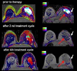
In these images of the breast, the lighter and brighter the color, the more aggressive the tumor and the greater the growth of angiogenesis, or the blood vessel growth around them. Functional MRI reveals small islands of the tumor that are resistant to chemotherapy <br>
Scientists are using new imaging technology to help them perform “virtual biopsies,” – biological profiles of specific tumors that may help predict a patient’s response to treatment and probability of long-term survival. This whole new realm of imaging is called functional MRI (magnetic resonance imaging), a process that offers insight into a tumor’s character, not just its superficial structure.
Using functional MRI, Dr. Michael Knopp, a radiologist and a member of The Ohio State University Comprehensive Cancer Center’s Experimental Therapeutics Program, is studying breast, prostate, pancreatic tumors and others to see if some of their particular biological quirks are related to response to treatment and survival.
Knopp says while X-rays can reveal information about a tumor’s size and shape, that information alone is not enough to help physicians plan and tailor some of the newest treatments. “It’s not what we see, but what we don’t that may be more important.”
What X-rays don’t show, but what functional MRI does, says Knopp, includes biological processes like angiogenesis, or blood vessel growth surrounding a tumor. Using MRI and special contrast agents, Knopp is able to determine the permeability, or “leakiness” of the tumor’s support system. Early studies suggest the “leakier” the vessels, the more likely a patient will respond to treatment. “Functional MRI allows us to measure permeability; understanding that characteristic alone can help clinicians better manage the patient’s care,” says Knopp.
Functional MRI can also reveal a tumor’s interior landscape, or it’s heterogeneity. Knopp says some tumors are extremely heterogeneous – meaning they are not biologically uniform. Instead, many may contain clusters of “hot spots,” clumps of cells that are biologically different and often resistant to treatment. “Functional MRI can help us identify those areas, understand their particular features, and hopefully, design targeted therapies for those specific sites,” says Knopp.
In functional MRI, images are made by measuring minute radio waves produced when hydrogen atoms in the body are trapped and vibrate within a magnetic field. The varying intensity of the signal reveals structural features and biological patterns illuminated by injected contrast agents.
“Analyzing data from those images can help us literally see where some chemotherapies are effective, and others are not. We know, for example, that in many cases, treatment with chemotherapy may kill 70 or 80 percent of a cancer, but the remaining tumor cells remain problematic. Now, we can find out exactly where those resistant areas are and we can be more selective and precise with additional treatment,” says Knopp. (See http://www.jamesline.com/output/breastimages.htm for an illustration.)
While functional MRI offers new ways to visualize cancer at work, it presents several problems that need to be solved before it becomes routinely useful in clinical care. It is still so new that scientists have yet to agree on standard methodology they will use to visualize what they want to see. That makes comparing studies and findings across multiple centers difficult. In addition, one study alone can generate as many as 700-800 images that need to be synthesized and read collectively for a complete analysis – a process requiring substantial computational power and highly-trained specialists.
“It’s an emerging field, and we think we are just beginning to see what it can do,” says Knopp.
Knopp reviewed functional MRI in oncology in an article in the April issue of Molecular Cancer Therapeutics.
His research is supported by the National Cancer Institute,The Wright Center of Innovation and the Ohio Biomedical Research and Technology Transfer Fund.
Media Contact
More Information:
http://www.osumedcenter.edu/All latest news from the category: Health and Medicine
This subject area encompasses research and studies in the field of human medicine.
Among the wide-ranging list of topics covered here are anesthesiology, anatomy, surgery, human genetics, hygiene and environmental medicine, internal medicine, neurology, pharmacology, physiology, urology and dental medicine.


Newest articles

Bringing bio-inspired robots to life
Nebraska researcher Eric Markvicka gets NSF CAREER Award to pursue manufacture of novel materials for soft robotics and stretchable electronics. Engineers are increasingly eager to develop robots that mimic the…

Bella moths use poison to attract mates
Scientists are closer to finding out how. Pyrrolizidine alkaloids are as bitter and toxic as they are hard to pronounce. They’re produced by several different types of plants and are…

AI tool creates ‘synthetic’ images of cells
…for enhanced microscopy analysis. Observing individual cells through microscopes can reveal a range of important cell biological phenomena that frequently play a role in human diseases, but the process of…

