Moving X-rays to revolutionise the diagnosis of back pain
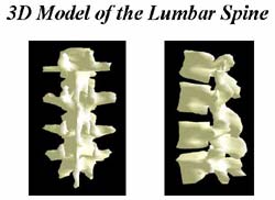
A ’solid model’ of the human lumbar spine
A new image processing system devised by engineers at the University of Southampton could change the way that back problems are diagnosed and provide a solution to one of the most common causes of work loss in the UK.
Low back pain is a significant problem and its cost to society is enormous. However, diagnosis of the underlying causes remains problematic despite extensive study. Reasons for this arise from the deep-rooted situation of the spine and also from its structural complexity.
Professor Robert Allen and his team in the Signal Processing & Control Group in the University’s Institute of Sound & Vibration Research are working with colleagues at the University’s Electronics & Computer Science department, The Anglo-European College of Chiropractic, and Salisbury Hospital, to develop a way of X-raying individuals while they are moving, a technique which they believe will improve the diagnosis of back problems by enabling clinicians to quantify how the spine is moving.
‘Up to now, clinicians have frequently used plain X-rays to diagnose back problems,’ comments Professor Allen. ‘These X-rays can only tell you about the spine in a static position, but if we X-ray as the person moves using very low dose radiation, we can see how the spine is moving and with image processing techniques, we can quantify the movement. Since back pain is often caused by soft tissue damage and not damage to the bones themselves, abnormal motion of the vertebrae may help us to locate the source of the problem.’
Their approach is based on automatically identifying vertebrae from the motion image sequences, calculating how each vertebrae moves and coupling this information with a dynamic 3-D lumbar spine visualisation. The traditional approach has been for clinicians to take 2-D images and to form a 3-D impression by mentally transforming these images. Three-dimensional visualisation of the lumbar spine can allow clinicians to observe the lumbar spine from different viewpoints and angles and may be helpful in understanding, diagnosing and treating back pain problems.
Their next challenge is to establish what ‘normal’ movement looks like so that they are in a position to recognise abnormalities.
Media Contact
All latest news from the category: Health and Medicine
This subject area encompasses research and studies in the field of human medicine.
Among the wide-ranging list of topics covered here are anesthesiology, anatomy, surgery, human genetics, hygiene and environmental medicine, internal medicine, neurology, pharmacology, physiology, urology and dental medicine.


Newest articles

Superradiant atoms could push the boundaries of how precisely time can be measured
Superradiant atoms can help us measure time more precisely than ever. In a new study, researchers from the University of Copenhagen present a new method for measuring the time interval,…

Ion thermoelectric conversion devices for near room temperature
The electrode sheet of the thermoelectric device consists of ionic hydrogel, which is sandwiched between the electrodes to form, and the Prussian blue on the electrode undergoes a redox reaction…

Zap Energy achieves 37-million-degree temperatures in a compact device
New publication reports record electron temperatures for a small-scale, sheared-flow-stabilized Z-pinch fusion device. In the nine decades since humans first produced fusion reactions, only a few fusion technologies have demonstrated…

