The breathing lifeline that comes at a price

However, the use of these machines can come at a price — every year thousands of patients are left with debilitating lung injuries, a small number of which are so serious the patient never recovers.
Now, a research collaboration between the universities of Nottingham and Leicester is to use computer modelling of lungs based on information collected from real patients to look at the best way of using ventilators to treat patients while minimising the risk of injury.
Dr Jonathan Hardman, of The University of Nottingham’s Division of Anaesthesia and Intensive Care, said: “For patients who don’t have the ability to breathe for themselves there is simply no other option than using a ventilator — it’s like carrying someone who is just too exhausted to walk.
“However, the use of these ventilators — which mechanically inflate and deflate the lungs — can cause tearing. You can be faced with a situation where a patient comes into the intensive care unit with a survivable illness but dies from a ventilator-associated injury. If they do make it out of the ICU, they could be left with lungs so badly scarred it could affect them for the rest of their life.
“Ventilator-associated injuries also extend the length of time a patient needs to spend in intensive care, putting them at risk of developing an un-related infection or the degradation of the muscles needed for breathing independently. In addition, these extra days spent on the ICU represent a huge cost to the NHS and affects the UK economy through loss of earnings from patients who are sick for longer than is necessary.
“We also have to count the human cost — it can be extremely distressing for families of patients to have to see their loved one supported by a ventilator.”
The challenge for researchers investigating ventilator-associated lung injury has been how to effectively monitor and observe the lungs of patients while on life-support. It is impossible to get monitoring equipment into the lungs themselves and x-rays are unable to provide the level of definition and clarity needed.
The £432,000 research project, funded by the Engineering and Physical Sciences Research Council (EPSCR) will see Dr Hardman working with control engineer Dr Declan Bates at The University of Leicester to produce believable computer models of lungs. These could be used to test a range of different uses of the ventilator, for example, varying the amount of oxygen supplied to the patient or the number of breaths per minute provided by the machine.
Real-life data collected in the autumn by researchers from patients on the Intensive Care Unit at Nottingham’s Queen’s Medical Centre will be used to create the computer models. The results of that work will then be taken out into clinical trials.
Dr Hardman said: “We plan to recreate a population of patients with a variety of illnesses and injuries which will allow us to look at the different permutations of treatment for those. Eventually this could lead to computer management of ventilators which will provide the optimum treatment with the least risk of injury.”
Media Contact
More Information:
http://www.nottingham.ac.ukAll latest news from the category: Health and Medicine
This subject area encompasses research and studies in the field of human medicine.
Among the wide-ranging list of topics covered here are anesthesiology, anatomy, surgery, human genetics, hygiene and environmental medicine, internal medicine, neurology, pharmacology, physiology, urology and dental medicine.
Newest articles

A new look at the consequences of light pollution
GAME 2024 begins its experiments in eight countries. Can artificial light at night harm marine algae and impair their important functions for coastal ecosystems? This year’s project of the training…

Silicon Carbide Innovation Alliance to drive industrial-scale semiconductor work
Known for its ability to withstand extreme environments and high voltages, silicon carbide (SiC) is a semiconducting material made up of silicon and carbon atoms arranged into crystals that is…

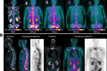
New SPECT/CT technique shows impressive biomarker identification
…offers increased access for prostate cancer patients. A novel SPECT/CT acquisition method can accurately detect radiopharmaceutical biodistribution in a convenient manner for prostate cancer patients, opening the door for more…


