Desminopathies: RUB researchers gain new insights into the mechanisms of heart disease
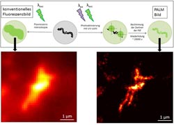
Using Photo Activation Localization Microscopy (PALM), ten times the resolution (right) of conventional light microscopy (left) is achieved. The image shows a desmin filament which was taken with a conventional microscope and with the high-resolution PALM microscope. Illustration: Andreas Brodehl/Per Niklas Hedde<br>
Malformed desmin proteins aggregate with intact proteins of the same kind, thereby triggering skeletal and cardiac muscle diseases, the desminopathies. This was discovered by researchers from the RUB Heart and Diabetes Center NRW in Bad Oeynhausen led by PD Dr. Hendrik Milting in an interdisciplinary research project with colleagues from the universities in Karlsruhe, Würzburg and Bielefeld. They report in the Journal of Biological Chemistry.
One defective gene is enough
Desmin normally forms stabilizing filaments inside of the cells. Different mutations in the DES gene, which contains the blueprint for the protein, induce different muscle diseases. Since chromosomes are always present in pairs, each cell has two DES genes on two different chromosomes. The desminopathies break out even if only one of the DES genes is mutated. With Photo Activation Localization Microscopy (PALM), the interdisciplinary team led by Dr. Milting revealed the mechanism behind this.
Making mutated and intact proteins visible
If one DES gene is mutated and one intact, a cell produces both malformed and normal proteins. Since not only the mutant desmin proteins clump together, but also the intact exemplars are incorporated into the aggregates, one defective DES gene is enough to trigger the disease. Using the PALM microscope, the researchers attach two different fluorescent molecules to the mutant and the intact proteins. They can turn these markers on and off by laser, effectively flashing them. From the “snapshots” of the intact and the mutated proteins, the computer then calculates a joint picture on which both protein variants can be seen. PALM is a novel microscopy technique that can achieve ten times higher resolution than conventional light microscopy.
Further research projects
In the next step, the research group would like to find out how mutations in the DES gene trigger what is termed arrhythmogenic right ventricular cardiomyopathy, ARVC for short. This rare heart muscle disease is characterized by a severe defect – especially to the right ventricle – and by heart rhythm problems that can lead to sudden cardiac death due to defects in the cell-cell contacts.
Bibliographic record
A. Brodehl et al. (2012): Dual-color photoactivation localization microscopy of cardiomyopathy associated desmin mutants, Journal of Biological Chemistry, doi: 10.1074/jbc.M111.313841
Further information
PD Dr. Hendrik Milting, Erich and Hanna Klessmann – Institute for Cardiovascular Research and Development, Heart and Diabetes Center NRW, Ruhr-Universität Bochum, Georgstraße 11, Bad Oeynhausen, Tel. 05731/97-3510
HMilting@hdz-nrw.de
Editor: Dr. Julia Weiler
Media Contact
More Information:
http://www.ruhr-uni-bochum.deAll latest news from the category: Health and Medicine
This subject area encompasses research and studies in the field of human medicine.
Among the wide-ranging list of topics covered here are anesthesiology, anatomy, surgery, human genetics, hygiene and environmental medicine, internal medicine, neurology, pharmacology, physiology, urology and dental medicine.


Newest articles

Bringing bio-inspired robots to life
Nebraska researcher Eric Markvicka gets NSF CAREER Award to pursue manufacture of novel materials for soft robotics and stretchable electronics. Engineers are increasingly eager to develop robots that mimic the…

Bella moths use poison to attract mates
Scientists are closer to finding out how. Pyrrolizidine alkaloids are as bitter and toxic as they are hard to pronounce. They’re produced by several different types of plants and are…

AI tool creates ‘synthetic’ images of cells
…for enhanced microscopy analysis. Observing individual cells through microscopes can reveal a range of important cell biological phenomena that frequently play a role in human diseases, but the process of…

