Unique image precision for disease treatment
Symbia IntevoTM*, the world's first xSPECT* system, integrates metabolic information from single photon emission computed tomography (SPECT) into computed tomography (CT) images.
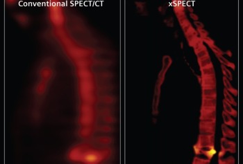
Previously, SPECT/CT images had low spatial resolution and physicians needed extensive experience and additional follow-up studies to decide whether a metabolic anomaly reflected a tumor or other diseases. Symbia Intevo also enables physicians to determine tumor size and thus plan treatment and monitor outcomes.
During SPECT examinations, patients are given low doses of radiopharmaceuticals that emit radiation when they react with a particular body tissue. Different metabolic processes can thus be observed depending on the agent administered. To determine the location of a metabolic disorder in the body, SPECT information is overlaid with CT images showing the anatomy of the body.
Until now, the problem has been that SPECT examinations offer only low spatial resolution and the high-precision CT images have to be adapted to match them. It can happen that the resulting image no longer clearly shows whether the metabolic disorder observed is inside or outside the bone. It would initially be unclear whether the anomaly was caused by a tumor in the bone or something else, such as a soft tissue inflammation.
The developers at Siemens Healthcare have now integrated SPECT and CT data in such a way that the high spatial resolution of the x-ray images remains intact and the SPECT images are significantly improved. The two datasets are generated sequentially during reconstruction in the same device using reference parameters such as the position of the detectors relative to the patient.
New, iterative image reconstruction algorithms refine the data in several passes. It was not previously possible to perform such complex calculation processes at the high resolution used in the CT images. That's why, in addition to new software, Symbia Intevo is also equipped with a powerful 64-bit computer.
The precise xSPECT data also makes it possible to determine the volume of the radiopharmaceutical used. This means that physicians can observe the change in metabolic activity and check whether their treatment is working.
Symbia Intevo also utilizes state-of-the-art algorithms that use the CT measurements to assign each voxel (three-dimensional pixel) in the xSPECT image to a particular class-fatty tissue, soft tissue, air, or hard (external) and soft (internal) bone areas. This makes it easy to recognize the body part where the metabolic disorder is located.
* Symbia Intevo and xSPECT are not commercially available in all countries. Due to regulatory reasons their future availability cannot be guaranteed.
Media Contact
All latest news from the category: Medical Engineering
The development of medical equipment, products and technical procedures is characterized by high research and development costs in a variety of fields related to the study of human medicine.
innovations-report provides informative and stimulating reports and articles on topics ranging from imaging processes, cell and tissue techniques, optical techniques, implants, orthopedic aids, clinical and medical office equipment, dialysis systems and x-ray/radiation monitoring devices to endoscopy, ultrasound, surgical techniques, and dental materials.


Newest articles

High-energy-density aqueous battery based on halogen multi-electron transfer
Traditional non-aqueous lithium-ion batteries have a high energy density, but their safety is compromised due to the flammable organic electrolytes they utilize. Aqueous batteries use water as the solvent for…

First-ever combined heart pump and pig kidney transplant
…gives new hope to patient with terminal illness. Surgeons at NYU Langone Health performed the first-ever combined mechanical heart pump and gene-edited pig kidney transplant surgery in a 54-year-old woman…

Biophysics: Testing how well biomarkers work
LMU researchers have developed a method to determine how reliably target proteins can be labeled using super-resolution fluorescence microscopy. Modern microscopy techniques make it possible to examine the inner workings…

