Ultra-high-field MRI may allow earlier diagnosis of Parkinson's disease
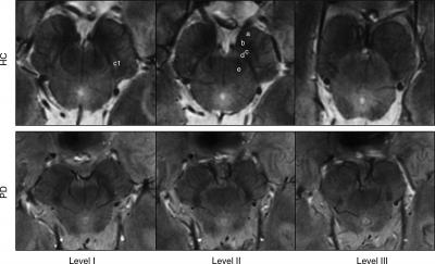
Top row: 7-T three-dimensional multiecho susceptibility-weighted in vivo images of SN in healthy 64-year-old man, located between the crus cerebri (a) and the red nucleus. Axial sections perpendicular to the floor of fourth ventricle are obtained at level of the inferior third of the red nucleus (level I), at the level of decussation of superior cerebellar peduncles (e) (level II), and at the level of the inferior colliculi (level III). At level I, SN appears as homogeneous hypointense structure in the medial part of the cerebral peduncle, and is laterally constituted by a hyperintense oval area between two hypointense layers (c1). At level II, a trilaminar organization of the SN with a central hyperintense layer (b) between two hypointense tiers (c and d) is detectable. At level III, the dorsal hypointense lamina could be detected as a small residual lateral hypointense area, while the hyperintense layer fades into the isointense cerebral peduncle. Bottom row: 7-T three-dimensional multiecho susceptibility-weighted in vivo images of the SN in PD patients. The loss of normal anatomy of the SN in a 61-year-old man with PD is characterized by the disappearance of the oval-shape bright spot in the lateral part of the SN at level I and by the loss of the hyperintense intermediate layer of the SN at level II. HC = healthy subject. Credit: Radiological Society of North America
Parkinson's disease is a chronic, progressive disease characterized by shaking, stiffness, and impaired balance and coordination. With no radiologic techniques available to aid in diagnosis, clinicians have had to rely on medical history and neurological examination. It is often difficult to distinguish Parkinson's disease from other conditions using these methods alone.
Mirco Cosottini, M.D., from the University of Pisa in Italy, and colleagues studied the brains of 38 individuals, including 17 Parkinson's disease patients and 21 healthy controls, as well as a brain specimen from a deceased individual, to help determine the accuracy of ultra-high-field 7-Tesla (7-T) MRI for identifying Parkinson's disease. Using the 7-T MRI, the researchers were able to distinguish a three-layered organization of the substantia nigra (SN), a crescent-shaped mass of cells in the midbrain.
Parkinson's disease results from the loss of dopamine-producing cells located in this region of the brain. Dopamine is an important neurotransmitter involved in multiple brain functions, including motor and behavioral processes such as mood, reward, addiction and stress. Based on abnormalities in the SN identified by the 7-T MRI, the researchers correctly classified patients with Parkinson's disease with a sensitivity of 100 percent and specificity of 96.2 percent.
According to Dr. Cosottini, the results show promise for earlier detection of the disease, which could speed the initiation of treatment.
“Parkinson's disease diagnosis remains clinically based, but with the introduction of 7-T MRI into clinical practice, a supporting radiologic diagnosis can be made,” he said.
The researchers also are exploring the clinical utility of 7-T MRI in several other neurodegenerative diseases, including mild cognitive impairment, a precursor of Alzheimer's disease.
“MR Imaging of the Substantia Nigra at 7 T Enables Diagnosis of Parkinson Disease.” Collaborating with Dr. Cosottini were Daniela Frosini, M.D., Ilaria Pesaresi, M.D., Mauro Costagli, Ph.D., Laura Biagi, Ph.D., Roberto Ceravolo, M.D., Ubaldo Bonuccelli, M.D., and Michela Tosetti, Ph.D.
Radiology is edited by Herbert Y. Kressel, M.D., Harvard Medical School, Boston, Mass., and owned and published by the Radiological Society of North America, Inc. (http://radiology.rsna.org/)
RSNA is an association of more than 53,000 radiologists, radiation oncologists, medical physicists and related scientists promoting excellence in patient care and health care delivery through education, research and technologic innovation. The Society is based in Oak Brook, Ill. (RSNA.org)
For patient-friendly information on MRI, visit RadiologyInfo.org.
Media Contact
All latest news from the category: Medical Engineering
The development of medical equipment, products and technical procedures is characterized by high research and development costs in a variety of fields related to the study of human medicine.
innovations-report provides informative and stimulating reports and articles on topics ranging from imaging processes, cell and tissue techniques, optical techniques, implants, orthopedic aids, clinical and medical office equipment, dialysis systems and x-ray/radiation monitoring devices to endoscopy, ultrasound, surgical techniques, and dental materials.


Newest articles

Silicon Carbide Innovation Alliance to drive industrial-scale semiconductor work
Known for its ability to withstand extreme environments and high voltages, silicon carbide (SiC) is a semiconducting material made up of silicon and carbon atoms arranged into crystals that is…

New SPECT/CT technique shows impressive biomarker identification
…offers increased access for prostate cancer patients. A novel SPECT/CT acquisition method can accurately detect radiopharmaceutical biodistribution in a convenient manner for prostate cancer patients, opening the door for more…

How 3D printers can give robots a soft touch
Soft skin coverings and touch sensors have emerged as a promising feature for robots that are both safer and more intuitive for human interaction, but they are expensive and difficult…

