New thoracic imaging approach can pinpoint underlying venous problems
Multi-detector computed tomography (CT) scanners are traditionally used to create three-dimensional images of arteries, the vessels which carry oxygen-rich blood away from the heart and distribute blood throughout the body. Veins, smaller vessels that return blood to the heart, are more difficult to accurately image.
Developed by Cristopher Meyer, MD and Achala Vagal, MD, the new protocol allows radiologists to compensate for the extra time it takes contrast solution to reach the veins so useful images can be produced using the CT scanner.
“We found that the rapid-imaging scanners were almost too fast for venous studies,” explains Vagal, a UC assistant professor and radiologist at University Hospital. “By the time the contrast reached the patient’s veins, there were too many artifacts to make any meaningful conclusions about possible disease—for example, blood clots.”
“Venous disease is rare and can be difficult to pinpoint,” she adds. “This new protocol uses the same imaging equipment in a novel way that allows us to acquire better venous images and make good clinical decisions.”
Vagal presented guidelines for this thoracic imaging protocol at the North American Society of Cardiovascular Imaging’s 35th Annual Meeting and Scientific Sessions in Washington, D.C., on Oct. 8.
For this new imaging technique, the CT technologist prepares two syringes of contrast: The first includes 140 cubic centimeters (CC) of undiluted contrast; the second contains a diluted mixture of 100 CC of contrast and 10 CC of saline solution.
“The key to getting accurate clinical images of the veins is in the timing,” Vagal says.
Both syringes are given consecutively at a rate of four CC per second, with a 60-second delay between the final injection and initiation of the CT scan.
“Previously, there was so much dense contrast in the veins that all you could see on the CT scan were streaks that didn’t tell you anything about possible venous disease,” explains Vagal. “Delaying the scan gave us enough time for both the arteries and the veins to be opacified, which resulted in the crisp images that allowed us to make better clinical determinations.”
Vagal is affiliated with the Neuroscience Institute at UC and University Hospital, a center of excellence that focuses on the main diseases of the brain and nerves such as stroke, brain tumors, brain trauma, Parkinson’s and Alzheimer’s disease, epilepsy, ALS and multiple sclerosis.
Media Contact
More Information:
http://www.uc.eduAll latest news from the category: Medical Engineering
The development of medical equipment, products and technical procedures is characterized by high research and development costs in a variety of fields related to the study of human medicine.
innovations-report provides informative and stimulating reports and articles on topics ranging from imaging processes, cell and tissue techniques, optical techniques, implants, orthopedic aids, clinical and medical office equipment, dialysis systems and x-ray/radiation monitoring devices to endoscopy, ultrasound, surgical techniques, and dental materials.


Newest articles
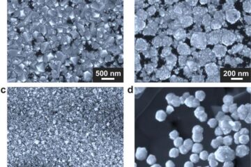
Making diamonds at ambient pressure
Scientists develop novel liquid metal alloy system to synthesize diamond under moderate conditions. Did you know that 99% of synthetic diamonds are currently produced using high-pressure and high-temperature (HPHT) methods?[2]…

Eruption of mega-magnetic star lights up nearby galaxy
Thanks to ESA satellites, an international team including UNIGE researchers has detected a giant eruption coming from a magnetar, an extremely magnetic neutron star. While ESA’s satellite INTEGRAL was observing…
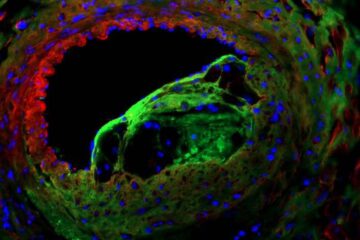
Solving the riddle of the sphingolipids in coronary artery disease
Weill Cornell Medicine investigators have uncovered a way to unleash in blood vessels the protective effects of a type of fat-related molecule known as a sphingolipid, suggesting a promising new…

