Three-dimensional, miniature endoscope opens new diagnostic possibilities
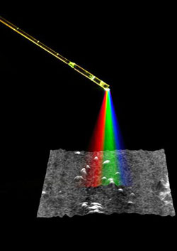
The spectral encoded miniature endoscope uses micro optics and a single optical fiber to project various colors of light onto different portions of the subject. The light reflected back into the endoscope is measured and analyzed to produce a three-dimensional image. This illustration shows a time exposure of white light transmitted through the miniature endoscope, superimposed on a three-dimensional rendering of mouse metastatic ovarian tumor nodules obtained with this new technique. Credit: Wellman Center for Photomedicine, Massachusetts General Hospital
Massachusetts General Hospital (MGH) researchers have developed a new type of miniature endoscope that produces three-dimensional, high-definition images, which may greatly expand the application of minimally invasive diagnostic and therapeutic procedures. In the October 19 issue of Nature, the team from the Wellman Center for Photomedicine at MGH describes their prototype device and a demonstration of its use in a mouse model.
“This new ultraminiature endoscope is the first to allow three-dimensional imaging of areas inside the body,” says Guillermo Tearney, MD, PhD, of the MGH Wellman Center, the report's senior author. “Its ability to go places that other imaging tools cannot reach opens new possibilities for medical diagnosis and eventually treatment.”
Standard miniature endoscopic devices – which give physicians access to hard-to-reach internal organs and structures – utilize bundles of optical fibers to supply light to and transmit images from the areas of interest. Larger endoscopes that use image sensors to produce high-quality, two-dimensional images can be a centimeter or more in diameter. Existing miniature endoscopes using smaller fiber bundles may be more flexible but have difficulty producing high-quality images.
The new device developed at MGH-Wellman uses a technology called spectrally encoded endoscopy (SEE). Multicolored light from a single optical fiber – introduced through a probe about the size of a human hair – is broken into its component colors and projected onto tissue, with each color illuminating a different part of the tissue surface. The light reflected back is recorded, and the intensity of the various colors decoded by a spectrometer, which analyzes the wavelengths of light. Another device called an interferometer, which calculates structural information based on the interaction between two waves of light, provides the data required to create three-dimensional images.
To demonstrate the device's application in a live animal, the researchers used the system to image metastatic ovarian tumors on the abdominal wall of a mouse. The SEE probe was passed into the abdominal cavity through a fine-gauge needle. The resulting three-dimensional image showed several raised areas of tumor nodules, the presence of which was confirmed by histologic analysis of the tissue.
“The most important feature of this new endoscope is the ability to obtain three-dimensional images, something we don't believe is offered by any commercially available miniature endoscope system,” says Dvir Yelin, PhD, first author of the Nature paper. “While the image resolution we achieved in this demonstration is similar to existing small-diameter endoscopes, with further optimization of the optics it is possible to obtain images with 10 times the number of pixels provided by other miniature endoscopes.”
“This new technology will offer physicians and surgeons the capability to bring many more procedures into outpatient settings, reduce anesthesia requirements and minimize tissue damage,” Tearney adds. “The device's size and flexibility should allow safer navigation through such delicate structures as the salivary ducts, the fallopian tubes and the pancreatic duct. Fetal and pediatric procedures may also benefit from this tool. Eventually, SEE could give rise to new procedures that permit diagnosis and microsurgery in previously inaccessible areas of the body.”
Media Contact
More Information:
http://www.mgh.harvard.edu/All latest news from the category: Medical Engineering
The development of medical equipment, products and technical procedures is characterized by high research and development costs in a variety of fields related to the study of human medicine.
innovations-report provides informative and stimulating reports and articles on topics ranging from imaging processes, cell and tissue techniques, optical techniques, implants, orthopedic aids, clinical and medical office equipment, dialysis systems and x-ray/radiation monitoring devices to endoscopy, ultrasound, surgical techniques, and dental materials.


Newest articles

“Nanostitches” enable lighter and tougher composite materials
In research that may lead to next-generation airplanes and spacecraft, MIT engineers used carbon nanotubes to prevent cracking in multilayered composites. To save on fuel and reduce aircraft emissions, engineers…

Trash to treasure
Researchers turn metal waste into catalyst for hydrogen. Scientists have found a way to transform metal waste into a highly efficient catalyst to make hydrogen from water, a discovery that…

Real-time detection of infectious disease viruses
… by searching for molecular fingerprinting. A research team consisting of Professor Kyoung-Duck Park and Taeyoung Moon and Huitae Joo, PhD candidates, from the Department of Physics at Pohang University…

