Cone beam CT proves better for visualizing some causes of hearing loss at half the radiation dose
The study, conducted in Bruges, Belgium, included 21 patients who had both a cone beam CT and a multidetector CT examination of their right and left temporal bones, said David Volders, MD, one of the authors of the study. Two radiologists reviewed the images from both exams and scored them based on image quality and the presence of pathology.
The study found that cone beam CT “corrected a false positive diagnosis for superior semicircular canal dehiscence in 11 out of 16 cases which were positive on multidetector CT (68.8%),” said Dr. Volders. Multidetector CT had indicated there was a dehiscence of the superior semicircular canal, when there wasn't, he said. In addition, cone beam CT scored significantly better than multidetector CT in visualizing normal temporal bone anatomy, said Dr. Volders.
“In our facility, all patients who undergo temporal bone imaging to diagnose fractures, congenital middle ear deformities, chronic ear infections and conductive hearing loss are now scanned with cone beam CT,” said Dr. Volders. “The significantly better image quality and the very low radiation dose has made cone beam CT our main choice for temporal bone imaging,” he said. “Radiologists should closely follow the cone beam CT evolutions and consider a cone beam CT in their practice as new generation high end cone beam CT is more and more claiming its place in diagnostic imaging of the temporal bone.” Dr. Volders added.
The study will be presented May 2, 2012 at the American Roentgen Ray Society Annual Meeting in Vancouver, Canada.
About ARRS
The American Roentgen Ray Society (ARRS) was founded in 1900 and is the oldest radiology society in the United States. Its monthly journal, the American Journal of Roentgenology, began publication in 1906. Radiologists from all over the world attend the ARRS Annual Meeting to take part in instructional courses, scientific paper presentations and scientific and commercial exhibits related to the field of radiology. The Society is named after the first Nobel Laureate in Physics, Wilhelm Röentgen, who discovered the X-ray in 1895.
Media Contact
More Information:
http://www.arrs.orgAll latest news from the category: Medical Engineering
The development of medical equipment, products and technical procedures is characterized by high research and development costs in a variety of fields related to the study of human medicine.
innovations-report provides informative and stimulating reports and articles on topics ranging from imaging processes, cell and tissue techniques, optical techniques, implants, orthopedic aids, clinical and medical office equipment, dialysis systems and x-ray/radiation monitoring devices to endoscopy, ultrasound, surgical techniques, and dental materials.


Newest articles
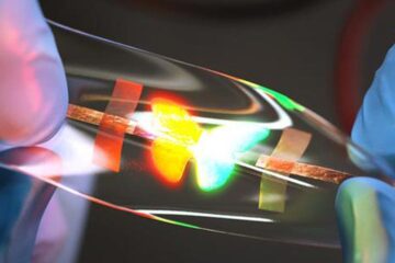
Stretchable quantum dot display
Intrinsically stretchable quantum dot-based light-emitting diodes achieved record-breaking performance. A team of South Korean scientists led by Professor KIM Dae-Hyeong of the Center for Nanoparticle Research within the Institute for…

Internet can achieve quantum speed with light saved as sound
Researchers at the University of Copenhagen’s Niels Bohr Institute have developed a new way to create quantum memory: A small drum can store data sent with light in its sonic…
A chip unique in the world
A team from UPV and iPRONICS has manufactured the first universal, programmable and multifunctional photonic chip on the market. A team from the Photonics Research Laboratory (PRL)-iTEAM of the Universitat…

