Same pieces, different picture – Unprecedented detail on HIV structure reveals surprises
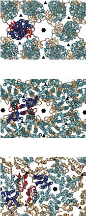
Surprisingly, the building blocks in immature HIV (centre) are arranged differently from those of immature Mason-Pfizer Monkey Virus (top). To form the mature virus, HIV’s building blocks take on yet another arrangement (bottom). Credit: EMBL/F.Schur
Scientists at the European Molecular Biology Laboratory (EMBL) in Heidelberg, Germany and collaborators from Heidelberg University, in the joint Molecular Medicine Partnership Unit, have obtained the first structure of the immature form of HIV at a high enough resolution to pinpoint exactly where each building block sits in the virus. The study, published online today in Nature, reveals that the building blocks of the immature form of HIV are arranged in a surprising way.
“The structure is definitely different from what we’d expected,” says John Briggs from EMBL, who led the work. “We assumed that retroviruses like HIV and Mason-Pfizer Monkey Virus would have similar structures, because they use such similar building blocks, but it turns out that their immature forms are surprisingly different from each other. At this point, we don’t really know why.”
Briggs and colleagues used cryo-electron microscopy to study the protein lattice that surrounds the virus’ genetic material. After infecting one of the cells in our immune system, HIV replicates, producing more copies of itself, each of which has to be assembled from a medley of viral and cellular components into an immature virus. This is the form that leaves the cell. The protein building blocks that make up the virus are then rearranged into the virus’ mature form, which can infect other cells.
The first cryo-electron microscopy images of immature HIV, obtained at EMBL in the 1990s, surprised researchers by showing that the virus did not have a regular symmetrical structure, as had been assumed. That meant it was going to be difficult to get a detailed picture of the structure of its protein lattice. Two decades on, by optimising both how data is collected at the microscope and how it is analysed, Florian Schur, a PhD student in Briggs’ lab, has now achieved an unprecedentedly detailed structure.
With this structure in hand, scientists have a basis to probe further. They can use it to decide where to focus efforts for achieving the even greater detail needed to explore potential drug targets, for instance. It will also enable researchers to understand how mutations might influence how the virus assembles. And the techniques themselves can be applied to a variety of questions.
“This approach offers so many possibilities,” says Schur. “You can look at other viruses, of course, but also at complexes and proteins inside cells, with a whole new level of detail.”
In future, the EMBL scientists will use the approach to look at other viruses and at the vesicles that transport material inside cells. They also aim to push the techniques even further, to allow them to see other parts of the viral proteins that are currently beyond their reach, but which they suspect play an important role in HIV maturation.
“In the long term, we’d also like to investigate how drugs which are known to inhibit virus assembly and maturation actually work,” Briggs concludes.
The study was conducted by the EMBL scientists together with their collaborators Barbara Müller and Hans-Georg Kräusslich at the University Clinic Heidelberg, in the joint Molecular Medicine Partnership Unit.
Published online in Nature on 22 October 2014.
Scientists at the European Molecular Biology Laboratory (EMBL) in Heidelberg, Germany and collaborators from Heidelberg University, in the joint Molecular Medicine Partnership Unit, have obtained the first structure of the immature form of HIV at a high enough resolution to pinpoint exactly where each building block sits in the virus. The study, published online today in Nature, reveals that the building blocks of the immature form of HIV are arranged in a surprising way.
“The structure is definitely different from what we’d expected,” says John Briggs from EMBL, who led the work. “We assumed that retroviruses like HIV and Mason-Pfizer Monkey Virus would have similar structures, because they use such similar building blocks, but it turns out that their immature forms are surprisingly different from each other. At this point, we don’t really know why.”
Briggs and colleagues used cryo-electron microscopy to study the protein lattice that surrounds the virus’ genetic material. After infecting one of the cells in our immune system, HIV replicates, producing more copies of itself, each of which has to be assembled from a medley of viral and cellular components into an immature virus. This is the form that leaves the cell. The protein building blocks that make up the virus are then rearranged into the virus’ mature form, which can infect other cells.
The first cryo-electron microscopy images of immature HIV, obtained at EMBL in the 1990s, surprised researchers by showing that the virus did not have a regular symmetrical structure, as had been assumed. That meant it was going to be difficult to get a detailed picture of the structure of its protein lattice. Two decades on, by optimising both how data is collected at the microscope and how it is analysed, Florian Schur, a PhD student in Briggs’ lab, has now achieved an unprecedentedly detailed structure.
With this structure in hand, scientists have a basis to probe further. They can use it to decide where to focus efforts for achieving the even greater detail needed to explore potential drug targets, for instance. It will also enable researchers to understand how mutations might influence how the virus assembles. And the techniques themselves can be applied to a variety of questions.
“This approach offers so many possibilities,” says Schur. “You can look at other viruses, of course, but also at complexes and proteins inside cells, with a whole new level of detail.”
In future, the EMBL scientists will use the approach to look at other viruses and at the vesicles that transport material inside cells. They also aim to push the techniques even further, to allow them to see other parts of the viral proteins that are currently beyond their reach, but which they suspect play an important role in HIV maturation.
“In the long term, we’d also like to investigate how drugs which are known to inhibit virus assembly and maturation actually work,” Briggs concludes.
The study was conducted by the EMBL scientists together with their collaborators Barbara Müller and Hans-Georg Kräusslich at the University Clinic Heidelberg, in the joint Molecular Medicine Partnership Unit.
Published online in Nature on 22 October 2014. DOI: 10.1038/nature13838
For images, video and more information please visit: www.embl.org/press/2014/141102_Heidelberg
Policy regarding use
EMBL press and picture releases including photographs, graphics and videos are copyrighted by EMBL. They may be freely reprinted and distributed for non-commercial use via print, broadcast and electronic
Sonia Furtado Neves
EMBL Press Officer & Deputy Head of Communications
Meyerhofstr. 1, 69117 Heidelberg, Germany
Tel.: +49 (0)6221 387 8263
Fax: +49 (0)6221 387 8525
sonia.furtado@embl.de
http://s.embl.org/press
For images, video and more information please visit: www.embl.org/press/2014/141102_Heidelberg
——————————
Policy regarding use
EMBL press and picture releases including photographs, graphics and videos are copyrighted by EMBL. They may be freely reprinted and distributed for non-commercial use via print, broadcast and electronic media, provided that proper attribution to authors, photographers and designers is made.
——————————
Sonia Furtado Neves
EMBL Press Officer & Deputy Head of Communications
Meyerhofstr. 1, 69117 Heidelberg, Germany
Tel.: +49 (0)6221 387 8263
Fax: +49 (0)6221 387 8525
sonia.furtado@embl.de
http://s.embl.org/press
Media Contact
All latest news from the category: Life Sciences and Chemistry
Articles and reports from the Life Sciences and chemistry area deal with applied and basic research into modern biology, chemistry and human medicine.
Valuable information can be found on a range of life sciences fields including bacteriology, biochemistry, bionics, bioinformatics, biophysics, biotechnology, genetics, geobotany, human biology, marine biology, microbiology, molecular biology, cellular biology, zoology, bioinorganic chemistry, microchemistry and environmental chemistry.


Newest articles

Silicon Carbide Innovation Alliance to drive industrial-scale semiconductor work
Known for its ability to withstand extreme environments and high voltages, silicon carbide (SiC) is a semiconducting material made up of silicon and carbon atoms arranged into crystals that is…

New SPECT/CT technique shows impressive biomarker identification
…offers increased access for prostate cancer patients. A novel SPECT/CT acquisition method can accurately detect radiopharmaceutical biodistribution in a convenient manner for prostate cancer patients, opening the door for more…

How 3D printers can give robots a soft touch
Soft skin coverings and touch sensors have emerged as a promising feature for robots that are both safer and more intuitive for human interaction, but they are expensive and difficult…

