Molecular motors may speed nutrient processing
At the time, Tyska knew that the core bundle traveling up the center of the microvillus was an array of the structural protein actin, and that the ladder-like “rungs” connecting the actin bundle to the cell membrane were composed of the motor protein myosin-1a. This myosin, though related to the myosin involved in muscle cell contraction, was thought to serve a purely structural role. “The textbook thinking for decades was that microvilli serve as a passive scaffold, a way to amplify the membrane surface area,” said Tyska, assistant professor of Cell and Developmental Biology at Vanderbilt University.
In the intestines, an expanded cell surface increases the space for nutrient-processing enzymes and transporters, offering greater capacity for nutrient handling. But it didn't make sense to Tyska that a motor protein – a protein with the potential to generate force and move cargo around in cells – would play a passive structural role. “When I looked at that image, the near crystalline arrangement reminded me of actin and myosin in a muscle fiber,” Tyska said. “I kept returning to the same question: why would the microvillus have this beautiful structure packed with motor proteins. The concentration of myosin motors in a single microvillus is very high; there’s serious force-generating potential there.”
Tyska and Russell McConnell, a student in his laboratory, tested the idea that these motor proteins are more than molecular glue binding the cell membrane to the actin bundle” The investigators purified the intestinal “brush border” – the layer of densely packed microvilli – from the intestines of rats or mice, and added ATP, the chemical fuel for myosin-1a. Through the microscope, they watched the cell membrane move toward the tips of the microvilli and pop off the ends in the form of vesicles, tiny bubble-like packets.
Their findings, reported in the May 21 Journal of Cell Biology with one of their images featured on the issue cover, have implications for nutrient processing and other aspects of gastrointestinal physiology. Tyska is excited about the group’s unexpected discovery. “What we’re showing is that the microvillus is more than just a scaffold to increase the amount of cell membrane,” Tyska said. “It’s a little machine that can shed membrane from the tips.” The team confirmed that myosin-1a is the motor that moves membrane up the microvillus. Brush borders isolated from knockout mice lacking the myosin-1a gene shed membrane at only five percent of the level of brush borders from wild-type animals.
The investigators are working now to understand why intestinal cells might launch vesicles from their microvilli. They know from ongoing vesicle sorting and mass spectrometry studies that the vesicles contain nutrient-processing enzymes and transporters, like the microvillar membrane. “One idea is that these vesicles operate remotely to speed nutrient processing, before the nutrients even get to the brush border to be absorbed by the (intestinal epithelial cell),” Tyska said.
The team is also exploring other possibilities for the role of membrane shedding: that it offers protection against microbes and pathogens by expelling them from the surface before they can enter the cell; that it provides a mechanism for altering the composition of the microvillar surface to handle changes in “what comes down the pipe;” and that it serves a role in cell-cell communication by launching vesicles that contain signaling proteins. Tyska and his team also plan to explore whether myosin-1a is serving a similar membrane-moving role in its other known location: the hair cells of the inner ear, and if other microvilli also use myosin motors to jettison vesicles from their tips.
Media Contact
More Information:
http://www.vanderbilt.eduAll latest news from the category: Life Sciences and Chemistry
Articles and reports from the Life Sciences and chemistry area deal with applied and basic research into modern biology, chemistry and human medicine.
Valuable information can be found on a range of life sciences fields including bacteriology, biochemistry, bionics, bioinformatics, biophysics, biotechnology, genetics, geobotany, human biology, marine biology, microbiology, molecular biology, cellular biology, zoology, bioinorganic chemistry, microchemistry and environmental chemistry.


Newest articles
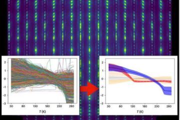
Machine learning algorithm reveals long-theorized glass phase in crystal
Scientists have found evidence of an elusive, glassy phase of matter that emerges when a crystal’s perfect internal pattern is disrupted. X-ray technology and machine learning converge to shed light…
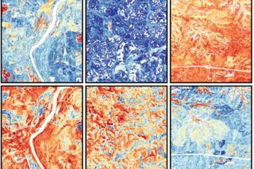
Mapping plant functional diversity from space
HKU ecologists revolutionize ecosystem monitoring with novel field-satellite integration. An international team of researchers, led by Professor Jin WU from the School of Biological Sciences at The University of Hong…
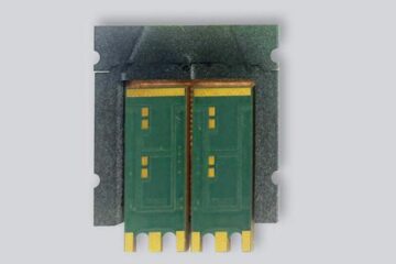
Inverters with constant full load capability
…enable an increase in the performance of electric drives. Overheating components significantly limit the performance of drivetrains in electric vehicles. Inverters in particular are subject to a high thermal load,…

