’Underground’ tunnels discovered as means for communication between immune system cells
University of Pittsburgh researchers first to report function of tunneling nanotubules
Immune system cells are connected to each other by an extensive network of tiny tunnels that, like a building’s hidden pneumatic tube system, are used to shoot signals to distant cells. This surprising discovery, being reported by two University of Pittsburgh School of Medicine researchers in the September issue of the journal Immunity, may explain how an immune response can be so exquisitely swift. The research not only proves cells other than neurons are capable of long-distance communication, but it reveals a hereto-unknown mechanism cells use for exchanging information.
Blood-derived dendritic cells and macrophages, both antigen-presenting cells, make use of these so-called tunneling nanotubules to relay molecular messages, report Simon C. Watkins, Ph.D., and Russell D. Salter, Ph.D. Further research may show there are additional cell types with these microscopic tunnel connections. Thus far, their studies suggest the tunnels do not exist between commonly used fibroblast and tumor cell lines.
Interestingly, if not for a minor mishap while carrying out an experiment, the authors might not have discovered the existence of these physical structures and conducted the studies that revealed their role in intercellular communication.
Using a custom-built, multi-camera live cell microscopic imaging system, they report that, in a matter of seconds, dendritic cells and macrophages can send waves of calcium and other small molecules to cells hundreds of micrometers away. Each nanotubule measures between 35 and 200 nanometers across – 5000 times smaller than the width of a human hair – and at any given time, cells may have up to 75 of these extensions, each of varying lengths.
“Considering their scale, these nanotubules are allowing communication between fairly distant cells. If instead of a culture dish we were talking about a large metropolitan area, the distance would be about the equivalent to four or five city blocks. That’s nothing short of amazing,” remarked Dr. Salter, associate professor of immunology at the University of Pittsburgh School of Medicine.
The authors are the first to explain the function of tunneling nanotubules, structures that were first described in fruit flies in 1998, and subsequently, identified in a handful of different types of animal and human cells.
“It’s one thing to find that this intricate physical network exists but quite astonishing to learn that immune system cells are using it to relay molecular signals to one another,” said Dr. Watkins, professor and vice chair, department of cell biology and physiology, and director of the Center for Biologic Imaging, University of Pittsburgh School of Medicine.
While gap junctions – interconnecting molecular bridges that conjoin tightly packed cells – are known to generate calcium signals and transport other molecules between cells, the researchers say the tunneling nanotubules are something quite different.
“This is clearly a third form of intercellular communication, distinct from gap junctions and synapses used by nerve cells. And, it is possible that tunneling nanotubules are essential for the function of the immune system, just as gap junctions are critical for the function of cardiac muscle. Exactly how this is so, we don’t know,” added Dr. Watkins, who also is a professor of immunology.
“Further study may help us better understand how they’re involved in the local inflammatory response of the immune system. For instance, we may find that dendritic cells use this network to distribute antigens to other cells and it may be conceivable to follow the entire pathway by tracing the network of tunneling nanotubules,” said Dr. Salter.
The authors’ discovery builds on their recent research showing how dendritic cells respond to stimuli, but, as they freely admit in this paper, it was due in large part to an accidental observation, that giving just the slightest poke to a single cell can set off a chain reaction whereby cell after cell discharges bursts of calcium.
In their earlier studies, they described how dendritic cells unfurl hidden veils – membranes that are so thin they can barely be imaged – and use these veils to move in on and capture their target. In the presence of E. coli, this occurs so rapidly and with such vigor that in accelerated time-lapse video, the cells appear more like a pack of wild animals feeding on a carcass.
But two things baffled the researchers. Dendritic cells extended their veils even before making physical contact with E. coli, yet macrophages, cells not normally picky about the antigens they engulf, were completely unresponsive to the bacteria. In order to understand how dendritic cells first sense the presence of an antigen and why the reaction is cell-specific, the authors decided to look at calcium flux, a well-recognized early measure of stimulation in numerous cell types. The use of a fluorescent dye, which allows direct measurement of calcium levels, would determine if calcium flux occurs before dendritic cells unfurl their veils.
With a microinjection tip, they squirted a mixture of E. coli fragments into a culture dish, and, indeed, one to two minutes before the appearance of the thin membranes, there were bursts of color indicative of calcium flux. Given their earlier results, the researchers anticipated that by repeating the experiment with macrophages there’d be no response. But as luck would have it, the microscopic bacteria sample somehow got clogged inside the tip, and before Dr. Watkins realized the need to pull away from the cell, he had already given it a jab.
“On the screen it looked like flash bulbs going off in a dark concert arena,” Dr. Salter recalled of that moment, when to both their great surprise the researchers witnessed how that little mishap had caused the macrophages to release bursts of calcium.
Returning to dendritic cells, they found that by giving a deliberate poke with an empty microinjection tip it caused the same reaction. But why some cells responded and others did not made Drs. Salter and Watkins wonder if there was some sort of physical structure connecting only those cells that discharged. A literature search turned up a handful of papers describing tunneling nanotubules, and further imaging using the highest magnification possible disclosed their presence in both the dendritic cells and macrophages.
In their most definitive experiment, the researchers placed dendritic cells, macrophages and a small amount of the E. coli mixture in the same culture dish. The dendritic cells, as would be expected, fluxed calcium in response to the E. coli. But a few seconds later, calcium could also be seen shooting through the tiny tunnels extending from dendritic cells to neighboring macrophages.
“This may solve some of the mystery of how a local stimulus directed at a very small number of cells can be amplified and result in a successful immune response,” explained Dr. Watkins.
“Quite possibly, the tunneling nanotubules enable a small number of dendritic cells with captured antigens to reach other dendritic cells in lymph nodes, increasing the number of these cells capable of stimulating T lymphocytes,” added Dr. Salter.
The finding that nanotubules play a role in sending molecular signals to other immune system cells calls into question the long-held belief that immune system cells talk to one another solely by secreting substances such as cytokines, the authors say. It now seems clear that intercellular communication is much more complicated. While it would be fascinating to see this interplay inside living tissue, detecting the tiny tubules in such a complex environment may be nearly impossible with current technology.
Media Contact
More Information:
http://www.upmc.eduAll latest news from the category: Life Sciences and Chemistry
Articles and reports from the Life Sciences and chemistry area deal with applied and basic research into modern biology, chemistry and human medicine.
Valuable information can be found on a range of life sciences fields including bacteriology, biochemistry, bionics, bioinformatics, biophysics, biotechnology, genetics, geobotany, human biology, marine biology, microbiology, molecular biology, cellular biology, zoology, bioinorganic chemistry, microchemistry and environmental chemistry.


Newest articles

How 3D printers can give robots a soft touch
Soft skin coverings and touch sensors have emerged as a promising feature for robots that are both safer and more intuitive for human interaction, but they are expensive and difficult…
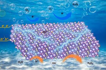
Oxygen vacancies mediated ultrathin Bi4O5Br2 nanosheets
… as efficient piezocatalyst for synthesis of H2O2 from pure water. As an important chemical raw material, hydrogen peroxide (H2O2) is widely applied in various aspects of industry and life….
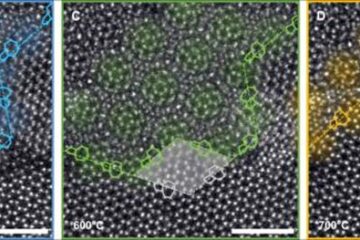
Atom-by-atom: Imaging structural transformations in 2D materials
Silicon-based electronics are approaching their physical limitations and new materials are needed to keep up with current technological demands. Two-dimensional (2D) materials have a rich array of properties, including superconductivity…

