Vision researchers find that photon receptors pair up in neat rows
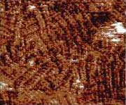
THE WELL-ORGANIZED EYE: This close up, high-magnification image of the disc membrane on a rod from the retina shows protrusions lined up in neat, double rows like eggs in a carton. The protrusions are a paracrystal form of rhodopsin, a light absorbing chemical. <br>
Using atomic-force microscopy, vision researchers have taken pictures of some of the eye’s photon receptors in their natural state, and have analyzed their packing arrangement. Their findings, published in the Jan. 9 issue of Nature, offer insight on how light signaling might be controlled in the retina’s outer edge.
The retina receives light through rods and cones. Rods, which are most heavily concentrated on the retina’s outer edge, are sensitive to dim light and to movement, but not to color. Rods, like cones, face away from incoming light. Within rods, light causes a chemical reaction with rhodopsin. This begins a chain of stimulation along the visual pathway, which sends information to the brain for interpretation. The brain can detect one photon of light, the smallest unit of energy, when it is absorbed by a photoreceptor.
The outer segment of a rod looks roughly like a stack of microscopic coins inside a wrapping. The segment is made up of discs covered by a membrane. Scientists studying the retina knew that the outer-segment disc membranes of rods are densely packed with rhodopsin molecules. This bunching together allows for optimum absorption of dim light and for subsequent amplification of the faint signal by the visual pathway. However, how rhodopsin molecules are physically arranged to increase the probability of being activated by a photon was not known.
In the Nature study, scientists looked at outer-segment disc membranes taken from rods in mouse retinae. Their collection method preserved the biological activity and organization of rhodopsin in the membranes. Atomic-force microscopy revealed that much of the surface of the membrane was markedly textured with narrow-ruled lines. At high magnification, researchers could see rhodopsin pairs appearing as tidy double rows of protrusions. They had the regularity of eggs in a carton.
Earlier, scientists conducting biochemical and pharmacological analyses had proposed that similar receptors were arranged in this manner. The recent demonstration that rhodopsin molecules pair in tight, neat lines is consistent with those inferences.
This particular organization of signaling molecules has important implications for recognition of particular proteins, binding, signal amplification, and signal termination. The shape and dimensions of the cell set stringent boundaries for this type of physical configuration, for contacts between the paired units, and for formation of larger structures consisting of these units.
The researchers on this study were Drs. Dimitrios Fotiadis and Andreas Engel of the M.E. Muller Institute for Microscopy, Biozentrum, University of Basel, Switzerland; and University of Washington researchers Drs. Yan Liang, Slawomir Filipek, and David Saperstein from the Department of Ophthalmology, and Dr. Krzysztof Palczewski, who is the Bishop Professor of Ophthalmology and also holds appointments in pharmacology and chemistry.
Media Contact
More Information:
http://www.washington.edu/All latest news from the category: Life Sciences and Chemistry
Articles and reports from the Life Sciences and chemistry area deal with applied and basic research into modern biology, chemistry and human medicine.
Valuable information can be found on a range of life sciences fields including bacteriology, biochemistry, bionics, bioinformatics, biophysics, biotechnology, genetics, geobotany, human biology, marine biology, microbiology, molecular biology, cellular biology, zoology, bioinorganic chemistry, microchemistry and environmental chemistry.


Newest articles

Silicon Carbide Innovation Alliance to drive industrial-scale semiconductor work
Known for its ability to withstand extreme environments and high voltages, silicon carbide (SiC) is a semiconducting material made up of silicon and carbon atoms arranged into crystals that is…

New SPECT/CT technique shows impressive biomarker identification
…offers increased access for prostate cancer patients. A novel SPECT/CT acquisition method can accurately detect radiopharmaceutical biodistribution in a convenient manner for prostate cancer patients, opening the door for more…

How 3D printers can give robots a soft touch
Soft skin coverings and touch sensors have emerged as a promising feature for robots that are both safer and more intuitive for human interaction, but they are expensive and difficult…

