Pioneering research on nuclear noncoding RNA

Shinichi Nakagawa
Initiative Research Scientist
Nakagawa Initiative Research Unit
RIKEN Advanced Science Institute
Research on the function of a type of RNA called ‘Gomafu’ is the primary field of study at RIKEN’s Nakagawa Initiative Research Unit. A central dogma in life science is that RNA, after being transcribed from genetic information in DNA in the nucleus of a cell, is transported out of the nucleus into the cytoplasm, where it is translated into proteins. In 2005, however, researchers were astonished to discover many RNAs that are not translated into proteins. These RNAs are referred to as ‘noncoding RNAs’. Gomafu RNA is one such noncoding type of RNA, and is unique in that it is not transported out of the nucleus. Why does Gomafu RNA stay in the nucleus, and how does it function? This article reports on revolutionary and pioneering research into nuclear noncoding RNAs.
WHY AM I HERE?
“In short, I want to know why I am here,” says Shinichi Nakagawa, Initiative Research Scientist and head of the Nakagawa Initiative Research Unit. “Mammals, and particularly humans, have a large and very complex nervous system. What is the mechanism behind the formation of the nervous system? I am working to elucidate this mechanism through studies of the retina.”
Why use the retina? “The retina constitutes part of the mammalian nervous system. Optical information captured by the eye is received by the retina, and from there is transmitted to the brain via the optic nerve. The retina consists of six types of nerve cell and one type of glial cell in a regular laminar structure. Other nervous systems generally have many more types of cell. Additionally, the retina permits easier manipulation and drug entry compared to the brain, which is protected by the skull. The eye is therefore easy to handle and is commonly used in a broad range of studies of the nervous system.”
Nakagawa previously investigated axon guidance mechanisms of the optic nerve as a postdoctoral fellow at the Department of Anatomy, University of Cambridge. However, Nakagawa also has another reason for selecting the retina for his research. “I saw a very beautiful retina during a practical course as an undergraduate student. The retina was wrapped in a coat of pigment cells. I removed the coat and the beautiful translucent retina appeared. It is important for me that the subject of my research is beautiful.”
THE SIGNALING MOLECULE Wnt PRESERVES RETINAL STEM CELL CHARACTERISTICS
“The retina exhibits a wonderful phenomenon that has long been known about,” says Nakagawa. When retinal tissue is cultured as dissociated cells, the laminar structure seen in the properly formed retina is not obtained. Many researchers have been working unsuccessfully for more than 30 years to identify the factors that induce correct laminar formation in vitro. Nakagawa had a breakthrough with this (Fig. 1). “I obtained a protein from a laboratory that was, by chance, located close to mine. I cultured dissociated retinal cells in a culture medium containing this protein, and a normal laminar structure was formed.”
Usually, the desired factor cannot be found without many painstaking steps, such as the addition of various proteins, the manipulation of genes, and extensive analyses of the culture broth ingredients. “I was prepared to climb a high mountain. But when I set out, I found my goal right in front of me. I was very lucky.”
Wnt, a secreted signaling molecule, was found to be involved in the formation of the laminar structure of retinal cells. “Retinal stem cells, cells that can differentiate into all the cell types found in the retina, undergo vigorous replication and division in the presence of Wnt. Dissociated cells do not arrange themselves into a laminar structure; new cells of the same type are produced from the retinal stem cells to form the laminar structure. Wnt probably functions to allow the retinal stem cells to retain their original characteristics.”
This important discovery has attracted considerable attention because the factor that serves to maintain the retinal stem cell characteristics had not previously been identified.
ENCOUNTERING GOMAFU RNA
One of the characteristics of the mammalian brain is the laminar structure found in the human cerebral cortex. Cells in each of these layers are of the same type. “The regular layered formation of cells in the retina and cerebral cortex is quite interesting. Although Wnt allows the retinal stem cells to retain their essential characteristics, it does not seem to have layer-forming activity. What, then, causes the cells to organize into laminar structures? I wanted to clarify this mechanism.” Subsequently, after working as a researcher at the RIKEN Center for Developmental Biology, Nakagawa established his Initiative Research Unit.
Nakagawa set out on a new strategy based on the assumption that the characteristics of cells differ depending on the layer in which they are present. Identifying the factor that determines the characteristics should lead to elucidation of the mechanism involved in formation of the laminar structure. “First, I attempted to find genes that are expressed exclusively in a particular type of retinal cell. There are many of these genes. On examining the intracellular distributions of their RNA, I was surprised to find one type of RNA that had a distribution distinct from the other types.”
RNA is the product of transcription of the genetic region of DNA. The class of RNA that is translated into amino acid sequences as a precursor to protein biosynthesis is known as messenger RNA (Fig. 2).
“RNA transcribed from DNA in the nucleus is transported out of the nucleus and is then translated into protein. Usually, RNA is transported out of the nucleus immediately after transcription, so we see the RNA signal in the cytoplasm but not in the nucleus. This is common knowledge for researchers who have examined gene expression.”
However, one type of RNA was found to exhibit unique behavior (Fig. 3). “The center of the cell was not empty, meaning that the RNA was present in the nucleus. I had examined the distributions of many RNA up to that time, and I had never come across an RNA with this unique distribution.”
The distribution of this unique RNA is reminiscent of the pattern of colors on the skin of the spotted seal, which is called gomafu azarashi in Japanese. Nakagawa therefore decided to name this RNA ‘Gomafu’. “A newly discovered gene is usually named with a term relevant to its function. However, as the function of this gene was unclear, I gave it a name based on the appearance of its distribution in the cell.” Later, Gomafu RNA was found to be strongly expressed in nerve cells, particularly in those having large cell bodies.
The primary research theme of Nakagawa’s laboratory is to elucidate the function of Gomafu RNA. “You may wonder how this fits into my research into the mechanism involved in forming the laminar structure, but researchers tend to want to examine anything wonderful they encounter. In addition, the distribution of Gomafu RNA is very beautiful, so I couldn’t help but study it.”
NONCODING RNA THAT STAYS IN THE NUCLEUS
The discovery of Gomafu RNA raises two important questions. The first is why this particular RNA is not transported out of the nucleus. Are there other types of RNA that, like Gomafu, do not pass out of the nucleus? “As far as I know, there are only four such RNAs: Xist, NEAT1, NEAT2 and Gomafu.”
Gomafu and the Xist, NEAT1 and NEAT2 RNAs (Fig. 2) are not translated into proteins and are thus referred to as ‘noncoding RNA’. In the past, it was thought that only 2% of all DNA was transcribed into RNA and then translated into proteins, the majority of which have no function. In 2005, however, Yoshihide Hayashizaki, project director at the RIKEN Genomic Sciences Center and now Director of the RIKEN Omics Science Center, and his colleagues wrote a paper in which it was concluded that up to 70% of DNA is transcribed into RNA, many types of which are noncoding RNA that are not translated into proteins. Some of the noncoding RNAs were also suggested to have specific functions. The paper attracted broad attention. A list of noncoding RNAs revealed by the base sequence of the mouse genome is already available in a publicly accessible database.
“We have examined the cellular distributions of several hundred noncoding RNAs registered with the database, and identified only four RNAs that can be described as bona fide nuclear noncoding RNAs based on evidence that they accumulate in the nucleus. These are Gomafu, Xist, NEAT1 and NEAT2. Out of the many noncoding RNAs, why is Gomafu RNA not transported out of nucleus?”
The second question concerns the function of Gomafu. Xist RNA has been studied for many years, and is now known to bind to chromosome X like a cover to suppress its functionality. Many reports are available on NEAT1 and NEAT2 RNA, which are expressed in large amounts, yet their functions remain to be clarified. Gomafu RNA is also expressed at high levels, but its function also remains unknown.
Elucidation of the function of Gomafu RNA will undoubtedly answer the question of why it remains in the nucleus. “The first step will be to make use of existing techniques,” says Nakagawa. An effective approach for such a study is to inhibit the function of Gomafu RNA and look for changes. In addition, although RNA is generally unstable, Gomafu RNA seems to form a stable structure by binding to a particular protein. Identifying the binding protein may provide a further clue as to the function of Gomafu RNA.
The reliance on existing techniques inevitably involves limitations, and it may be impossible to elucidate the function of Gomafu RNA without introducing new analytical and experimental techniques. “A big advantage of RIKEN is that laboratories for a broad range of biological and other fields are available at one’s elbow. So, on reaching a deadlock in determining which approach to take, it is easy to ask an expert in a different field for hints on solving the problem,” continues Nakagawa.
Nakagawa took note of a paper produced as part of a joint research project between Toshihiro Tanaka, group director at the RIKEN Center for Genomic Medicine, and an Osaka University group, in which it was reported that the substitution of a single base in the MIAT gene with another particular base increases the probability of the onset of myocardial infarction by a factor of 1.38. MIAT and Gomafu RNA correspond to the same gene. Although Tanaka and his colleagues discovered the gene earlier, Nakagawa made his discovery before MIAT had been registered in the database, so the gene has been given two names. “I read the paper by Tanaka and his colleagues and became confident that the Gomafu RNA does something important. I was encouraged to continue my research into Gomafu.”
BEAUTIFUL WORLD UNDER THE MICROSCOPE
A visit to the Research Unit’s homepage reveals Nakagawa’s words of self-introduction: “I am pursuing the study of impressive things that can be seen under the microscope.” He says with a laugh, “I am fond of visually impressive phenomena. I feel happiest when I can see beautiful cells or tissue under the microscope. Director Hayashizaki and his colleagues were the first to register the base sequence of Gomafu RNA in the database. However, it was certainly me who observed its distribution under the microscope for the first time. I have never forgotten that moment.”
Nakagawa thinks it will not be easy to elucidate the function of Gomafu RNA. “When I reach the bottom of a slope on a bicycle, I cannot help but keep cycling up the hill. I feel no interest in doing something that is likely to be fully understood in a few years. It is clear that embarking on something that will take 20 or 30 years will pioneer new worlds.”
Ever since he was a child, Nakagawa wanted to become a scientist. He was profoundly impressed by two books he read while a junior high school student: Cellular Society – Exploring the Fundamentals of Biological Order by Tokindo Okada (Professor Emeritus at Kyoto University), and King Solomon’s Ring: New Light on Animals’ Ways by Konrad Lorenz. He subsequently entered Kyoto University with the aim of conducting research in biology. Although he entered the University’s Faculty of Agriculture, he moved to the Faculty of Science in his third year upon realizing that Okada’s laboratory was part of Keio University. He joined the laboratory of Masatoshi Takeichi (now Director of the RIKEN Center for Developmental Biology), who was the direct successor to Okada. “I am very proud that I was able to study in the laboratory run by Drs Okada and Takeichi.”
When asked about his dream as a researcher, Nakagawa answers, “I want to create a new field. Textbooks in biology have different chapters. I will be happy if I can write a new chapter entitled ‘Nuclear Noncoding RNA’.”
About the researcher
Shinichi Nakagawa was born in Suwa, Japan, in 1971. He graduated from the Faculty of Sciences, University of Kyoto, in 1993, and obtained his PhD in 1998 from the same university. After two years postdoctoral training at the Department of Anatomy, University of Cambridge, UK, he returned to Japan as Assistant Professor at Kyoto University. He then joined the RIKEN Center for Developmental Biology in Kobe, as a researcher, and later the RIKEN Advanced Science Institute in Wako as Initiative Researcher, where he started his career in developmental neurobiology. His research focuses on the molecular machinery regulating the physiological function of the nervous system.
Media Contact
All latest news from the category: Life Sciences and Chemistry
Articles and reports from the Life Sciences and chemistry area deal with applied and basic research into modern biology, chemistry and human medicine.
Valuable information can be found on a range of life sciences fields including bacteriology, biochemistry, bionics, bioinformatics, biophysics, biotechnology, genetics, geobotany, human biology, marine biology, microbiology, molecular biology, cellular biology, zoology, bioinorganic chemistry, microchemistry and environmental chemistry.
Newest articles

Properties of new materials for microchips
… can now be measured well. Reseachers of Delft University of Technology demonstrated measuring performance properties of ultrathin silicon membranes. Making ever smaller and more powerful chips requires new ultrathin…

Floating solar’s potential
… to support sustainable development by addressing climate, water, and energy goals holistically. A new study published this week in Nature Energy raises the potential for floating solar photovoltaics (FPV)…

Skyrmions move at record speeds
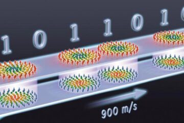
… a step towards the computing of the future. An international research team led by scientists from the CNRS1 has discovered that the magnetic nanobubbles2 known as skyrmions can be…


