New microscopy system captures 'lost' fluorescence, improving resolution
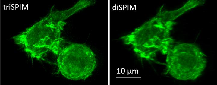
This is a macrophage actin labeled with green fluorescent protein, imaged with the new triSPIM microscope (left) and the diSPIM (right). Credit: Yicong Wu and Valentin Jaumouille
The researchers, who work collaboratively at the Marine Biological Laboratory's Whitman Center, published their results this week in the journal Optica.
“Everybody knows fluorescence imaging is inefficient in that the microscope only captures a portion of the light (spewing off the specimen),” says senior author Hari Shroff of the National Institute of Biomedical Imaging and Bioengineering. “In this paper, we showed you can not only capture that lost light, but use computation to fuse it to the existing image and make the image sharper.”
Developed by Yicong Wu, a staff scientist in Shroff's lab, the new system achieved resolution of up to 235 x 235 x 340 nanometers, which is double the volumetric resolution of traditional fluorescence microscopy methods.
To collect more of the available light (which, in turn, provides more information about the specimen), the new microscope has three objective lenses acquiring views of the sample simultaneously. The views are then aligned and merged by a computational process known as deconvolution.
Those computations were worked out in collaboration with co-author Patrick La Rivière of the University of Chicago's Radiology Department, who typically develops algorithms for improving “dose efficiency” in human-scale medical imaging, such as CAT scans.
“In medical imaging, we are always worried about dose, about capturing every X-ray [used on the patient to improve scan resolution]. We are concerned with 'How can we do more with less?'” La Rivière says.
In microscopy, the amount of light used presents similar concerns. “If you use very intense illuminations to image something microscopic like a worm embryo, you might change its biology or even kill it. You need to be dose efficient with your light,” La Rivière says.
La Rivière and Shroff began collaborating at the MBL in 2014, initially on algorithms to improve Shroff's diSPIM microscope (which has two objective lenses) and eventually on the new three-lensed microscope (called triSPIM).
La Rivière this year was named an MBL Fellow. Shroff is an MBL Whitman Center Scientist and co-director of the MBL's Optical Microscopy and Imaging in the Biomedical Sciences course.
###
Citation:
Yicong Wu, P. Chandris, P.W. Winter, E.Y. Kim, V. Jaumouillé, A. Kumar, M. Guo, J.M. Leung, C. Smith, I. Rey-Suarez, H. Liu, C.M. Waterman, K.S. Ramamurthi, P. La Riviere, H. Shroff (2016) Simultaneous multi-view capture and fusion improves spatial resolution in wide-field and light-sheet microscopy. Optica 3, 8: 897-920; doi: 10.1364/OPTICA.3.000897
The Marine Biological Laboratory (MBL) is dedicated to scientific discovery – exploring fundamental biology, understanding marine biodiversity and the environment, and informing the human condition through research and education. Founded in Woods Hole, Massachusetts in 1888, the MBL is a private, nonprofit institution and an affiliate of the University of Chicago.
Media Contact
All latest news from the category: Life Sciences and Chemistry
Articles and reports from the Life Sciences and chemistry area deal with applied and basic research into modern biology, chemistry and human medicine.
Valuable information can be found on a range of life sciences fields including bacteriology, biochemistry, bionics, bioinformatics, biophysics, biotechnology, genetics, geobotany, human biology, marine biology, microbiology, molecular biology, cellular biology, zoology, bioinorganic chemistry, microchemistry and environmental chemistry.


Newest articles

Superradiant atoms could push the boundaries of how precisely time can be measured
Superradiant atoms can help us measure time more precisely than ever. In a new study, researchers from the University of Copenhagen present a new method for measuring the time interval,…

Ion thermoelectric conversion devices for near room temperature
The electrode sheet of the thermoelectric device consists of ionic hydrogel, which is sandwiched between the electrodes to form, and the Prussian blue on the electrode undergoes a redox reaction…

Zap Energy achieves 37-million-degree temperatures in a compact device
New publication reports record electron temperatures for a small-scale, sheared-flow-stabilized Z-pinch fusion device. In the nine decades since humans first produced fusion reactions, only a few fusion technologies have demonstrated…

