First Images from Medical Beamline at Canadian Light Source
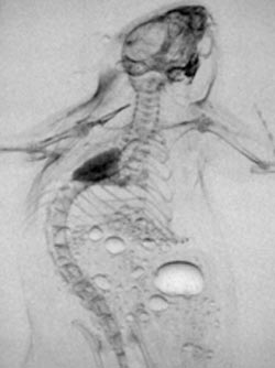
X-ray images of a mouse taken using a technique called diffraction-enhanced imaging (DEI).<br>Canadian Light Source
“Our entire team was just thrilled with what we saw – the images we’re getting on BMIT are as good as any I’ve gotten at any other synchrotron. That’s quite an achievement given that we had our first X-rays down the beamline less than a month ago,” said Dean Chapman, U of S Canada Research Chair in X-ray Imaging and BMIT team leader.
“The BMIT team is profoundly grateful to the staff of the CLS who have worked extremely hard over the past several years to bring this facility to fruition. In the coming months, we’ll be working closely with the CLS, the U of S and the Saskatoon Health Region to build the local, national and international research capacity of this world-class facility.”
“This is great news, both for the BMIT team and the Canadian Light Source,” said CLS Executive Director Josef Hormes. “BMIT is such an important facility, not just for the CLS but for the future investigation and treatment of diseases that have an impact on Canadians and people around the world.”
The X-ray images of a mouse were taken using a technique called diffraction-enhanced imaging (DEI), co-discovered by Chapman, U of S professor and former CLS Executive Director William Thomlinson, and U.S. researcher Zhong Zhong. The imaging team included Chapman, Thomlinson, CLS staff scientist Tomasz Wysokinski, U of S researcher David Cooper, graduate students Brian Bewer and Ying Zhu, and Dr. Sheldon Wiebe with the Saskatoon Health Region.
DEI uses the unique properties of synchrotron X-rays to produce images of soft tissues such as muscle, organs and tumours that do not readily absorb X-rays, making them cloudy or invisible to conventional X-ray radiographs and mammograms. For example, the mouse’s lungs are clearly visible in the DEI image, but only appear as a faint blur in the conventional absorption image.
DEI imaging is proving to be a valuable tool in visualizing cancer, imaging bone cartilage and understanding the structure and function of reproductive organs. Other techniques planned for development at BMIT include the delivery of precise beams of high energy X-rays for the treatment of cancer.
Under construction since fall 2005, BMIT is the only facility of its kind in North America and one of a handful in the world. The $17-million lab, consisting of two beamlines and support systems, will be complete in 2009. BMIT will be capable of imaging and therapy research involving plants and animals of all sizes – from hamsters to horses – and one day humans. The project is the result of an international collaboration of researchers from Canada, the U.S., Australia, France and Italy.
The Canadian Light Source is Canada’s national centre for synchrotron research. Located at the University of Saskatchewan in Saskatoon, the CLS is a powerful tool for academic and industrial research in a wide variety of areas including environmental science, natural resources and energy, health and life sciences, and information and communications technology. CLS operations are funded by the Government of Canada, NSERC, NRC, CIHR, the Government of Saskatchewan and the University of Saskatchewan.
Media Contact
All latest news from the category: Life Sciences and Chemistry
Articles and reports from the Life Sciences and chemistry area deal with applied and basic research into modern biology, chemistry and human medicine.
Valuable information can be found on a range of life sciences fields including bacteriology, biochemistry, bionics, bioinformatics, biophysics, biotechnology, genetics, geobotany, human biology, marine biology, microbiology, molecular biology, cellular biology, zoology, bioinorganic chemistry, microchemistry and environmental chemistry.


Newest articles

Superradiant atoms could push the boundaries of how precisely time can be measured
Superradiant atoms can help us measure time more precisely than ever. In a new study, researchers from the University of Copenhagen present a new method for measuring the time interval,…

Ion thermoelectric conversion devices for near room temperature
The electrode sheet of the thermoelectric device consists of ionic hydrogel, which is sandwiched between the electrodes to form, and the Prussian blue on the electrode undergoes a redox reaction…

Zap Energy achieves 37-million-degree temperatures in a compact device
New publication reports record electron temperatures for a small-scale, sheared-flow-stabilized Z-pinch fusion device. In the nine decades since humans first produced fusion reactions, only a few fusion technologies have demonstrated…

