Functional Magnetic Resonance Imaging under the Magnifying Glass
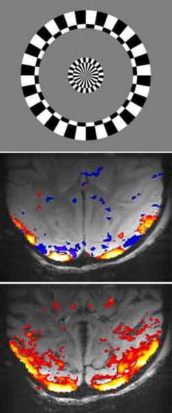
A: The stimulus used to elicit positive and negative BOLD responses in the visual cortex;<br>B: Positive and negative BOLD responses in monkey primary visual cortex to a visual stimulus;<br>C: The cerebral blood volume (CBV) response to a visual stimulus is increased in the entire primary visual cortex.<br><br>Jozien Goense / Max Planck Institute for Biologische Cybernetics, Tübingen<br>
The cortex consists of six different layers, which vary in their anatomical and physiological properties. It plays a key role in the cognitive capacities of the brain. Since the cortical layers are segregated functionally, we could potentially say something about the neural processes that take place when an area is activated if we could see different signals in the different layers.
Jozien Goense from the Max Planck Institute for Biological Cybernetics in Tübingen, Germany and her colleagues used functional Magnetic Resonance Imaging (fMRI) to observe these layer-specific neural processes within the cortex and found different mechanisms for fMRI response increases and decreases as well as cortical layer-dependent differences in the neurovascular coupling mechanism.
The cortex is the outermost layer of the brain and plays a key role in perception, memory, attention, thought, language and consciousness. In mammals, it consists of six horizontal layers, each with different anatomical and physiological properties and different connectivity. These layers have so far been elusive to study in vivo, for several reasons, like a lack of spatial resolution in functional Magnetic Resonance Imaging (fMRI), the inability to see deeper layers with various optical methods, or difficulty in determining the exact recording depth of electrodes. Therefore, if we can visualize the signals in the different layers, it would allow us to better probe the cortical circuitry, for example to determine the processing steps that occur between the input and output of a given cortical area.
Functional Magnetic Resonance Imaging (fMRI) is one of the most used tools to observe the functional activity of the brain. fMRI is a non-invasive method that measures brain activity by detecting associated changes in blood flow and oxygen consumption. The primary form of fMRI uses the blood-oxygenation-level-dependent (BOLD) contrast, which reflects the oxygen concentration in the blood, and through this indicates which brain areas are activated upon a certain stimulus. However, typical fMRI studies measure activation on the scale of a few millimeters and are not able to resolve the cortical layers. Furthermore, it is also not yet known if and how layer-specific neural activity is reflected in the BOLD-response. Other functional imaging methods that are less commonly used, but can shed light on this question, are based on the cerebral blood volume (CBV), whereby the amount of blood in the activated brain region is measured, or based on cerebral blood flow (CBF). These various methods have different sensitivities and measure different aspects of the blood flow response upon neural activity.
Jozien Goense is a project leader in the Department for Physiology of Cognitive Processes headed by Nikos Logothetis at the Max Planck Institute for Biological Cybernetics in Tübingen, Germany. She and her colleague Hellmut Merkle from the Laboratory of Functional and Molecular Imaging at the National Institutes of Health in Bethesda (USA), used high-resolution fMRI to measure BOLD-, CBV- and CBF responses to stimuli that elicit positive- and negative BOLD signals in the macaque primary visual cortex. They compared the activity patterns in response to excitatory stimuli, and stimuli that are known to give negative BOLD responses. Negative BOLD responses are reductions in the BOLD signal, often seen adjacent to stimulated regions. The negative BOLD signal is therefore thought to result from neuronal suppression.
They found that a negative BOLD response is not just the inverse of the positive response, but that it has a separate mechanism. Furthermore, the different layers responded differently to the stimuli. This indicates that the neurovascular coupling mechanism, which is the mechanism that provides the link between the neural signals and the BOLD-response, differs in the different layers and for the two stimuli. This means that potentially the layer-specific differences in the responses can be used to separate what kind of processes occur in the cortex.
These findings suggest different mechanisms for neurovascular coupling for BOLD increases and decreases as well as laminar differences in neurovascular coupling. The consequences of these findings are quite fundamental, since it may improve the interpretation of the BOLD signals in fMRI studies, and especially the negative one. Furthermore, it opens up the possibility to study neural processes within the cortical sheet, which would expand the applicability of fMRI and push it to smaller spatial scales than the ones it is currently used at.
Original Publication:
J. Goense, H. Merkle, N. K. Logothetis. (2012) High-resolution fMRI reveals laminar differences in neurovascular coupling between positive and negative BOLD responses. Neuron, doi: 10.1016/j.neuron.2012.09.019
Contact:
Dr. Jozien Goense
Phone: +49 7071 601-1704
E-mail: jozien.goense@tuebingen.mpg.de
Stephanie Bertenbreiter (Public Relations)
Phone: +49 7071 601-1792
E-mail: presse-kyb@tuebingen.mpg.de
The Max Planck Institute for Biological Cybernetics works in the elucidation of cognitive processes. It employs about 300 people from more than 40 countries and is located at the Max Planck Campus in Tübingen, Germany. The Max Planck Institute for Biological Cybernetics is one of 80 research institutes that the Max Planck Society for the Advancement of Science maintains in Germany and abroad.
Media Contact
All latest news from the category: Life Sciences and Chemistry
Articles and reports from the Life Sciences and chemistry area deal with applied and basic research into modern biology, chemistry and human medicine.
Valuable information can be found on a range of life sciences fields including bacteriology, biochemistry, bionics, bioinformatics, biophysics, biotechnology, genetics, geobotany, human biology, marine biology, microbiology, molecular biology, cellular biology, zoology, bioinorganic chemistry, microchemistry and environmental chemistry.


Newest articles

“Nanostitches” enable lighter and tougher composite materials
In research that may lead to next-generation airplanes and spacecraft, MIT engineers used carbon nanotubes to prevent cracking in multilayered composites. To save on fuel and reduce aircraft emissions, engineers…

Trash to treasure
Researchers turn metal waste into catalyst for hydrogen. Scientists have found a way to transform metal waste into a highly efficient catalyst to make hydrogen from water, a discovery that…

Real-time detection of infectious disease viruses
… by searching for molecular fingerprinting. A research team consisting of Professor Kyoung-Duck Park and Taeyoung Moon and Huitae Joo, PhD candidates, from the Department of Physics at Pohang University…

