F is for Fluoresence and Fluorine
The imaging of living cells at the molecular level was barely a dream twenty years ago. Today, however, this dream is close to becoming reality. In the Max Planck Institute for NanoBiophotonics in Göttingen, Stefan Hell (2009 recipient of the Otto Hahn Prize) has developed fluorescence microscopy methods for observing objects on the nanoscale and with his colleagues Vladimir Belov and Christian Eggeling a new series of photostable dyes that can be used as fluorescent markers has been realised, as reported in a cover story in Chemistry—A European Journal.
Over the last two decades Stefan Hell and his group have revolutionized the art of microscopy beyond limits thought to have been unbreakable. Due to the wave properties (diffraction) of light, the resolution of an optical microscope is limited to object details of about 0.2 micrometers. The laws of physics appeared to prohibit imaging details beyond this limit. Stefan Hell saw beyond this limitation and about fifteen years ago his vision became concrete; he developed a method for observing objects at the nanometer scale by sequentially turning the fluorescence of nearby molecules off by stimulated emission, a technique known as STED nanoscopy.
The sensitivity of this technique depends on the brightness of the applied fluorescence markers and their photostability is also of great importance. The NanoBiophotonics group has succeeded in synthesizing a series of highly photostable and highly fluorescent dyes. These compounds emit green and orange light and are based on fluorine derivatives of the well-known Rhodamine dye. The use of these dyes in STED nanoscopy leads to images of high-quality with respect to brightness and signal-to-background ratio; further the resolution over that of more traditional optical microscopes is significantly improved giving more detailed structural information.
These rhodamine-based fluorine derivatives are even more special because of their versatility. The compounds are available in hydrophilic and lipophilic forms, and with the inclusion of amino reactive groups, they can be easily attached to antibodies or other biomolecules in the course of standard labeling and immunostaining procedures. The group demonstrate that these new dyes are able to cross cellular membranes and reach the interior of living cells, which could lead to new labeling strategies for biological systems. All eyes are now on Göttingen to see just how far optical nanoscopy can go.
Author: Stefan Hell, Max Planck Institute of NanoBiophotonics, Göttingen (Germany), http://www.mpibpc.mpg.de/groups/hell/
Title: New Fluorinated Rhodamines for Optical Microscopy and Nanoscopy
Chemistry – A European Journal 2010, 16, No. 15, 4477–4488, Permalink to the article: http://dx.doi.org/10.1002/chem.200903272
Media Contact
All latest news from the category: Life Sciences and Chemistry
Articles and reports from the Life Sciences and chemistry area deal with applied and basic research into modern biology, chemistry and human medicine.
Valuable information can be found on a range of life sciences fields including bacteriology, biochemistry, bionics, bioinformatics, biophysics, biotechnology, genetics, geobotany, human biology, marine biology, microbiology, molecular biology, cellular biology, zoology, bioinorganic chemistry, microchemistry and environmental chemistry.


Newest articles
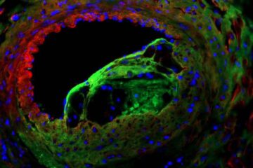
Solving the riddle of the sphingolipids in coronary artery disease
Weill Cornell Medicine investigators have uncovered a way to unleash in blood vessels the protective effects of a type of fat-related molecule known as a sphingolipid, suggesting a promising new…

Rocks with the oldest evidence yet of Earth’s magnetic field
The 3.7 billion-year-old rocks may extend the magnetic field’s age by 200 million years. Geologists at MIT and Oxford University have uncovered ancient rocks in Greenland that bear the oldest…
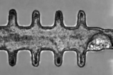
Mini-colons revolutionize colorectal cancer research
As our battle against cancer rages on, the quest for more sophisticated and realistic models to study tumor development has never been more critical. Until now, research has relied on…

