How to build your gate
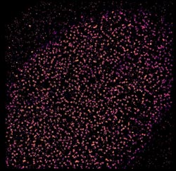
The Universe within: gates to the genome.<br>Credit: EMBL/A. Szymborska<br>
It's a parent's nightmare: opening a Lego set and being faced with 500 pieces, but no instructions on how to assemble them into the majestic castle shown on the box. Thanks to a new approach by scientists at the European Molecular Biology Laboratory (EMBL) in Heidelberg, Germany, researchers studying large sets of molecules with vital roles inside our cells can now overcome a similar problem. In a study published online today in Science, the scientists used super-resolution microscopy to solve a decade-long debate about the structure of the nuclear pore complex, which controls access to the genome by acting as a gate into the cell’s nucleus.
Like the flummoxed parent staring at the image on the box, scientists knew the gate’s overall shape, from electron tomography studies. And thanks to techniques like X-ray crystallography and single particle electron microscopy, they knew that the ring which studs the nucleus’ wall and controls what passes in and out is formed by sixteen or thirty-two copies of a Y-shaped building block. They even knew that each Y is formed by nine proteins. But how the Ys are arranged to form a ring was up for debate.
“When we looked at our images, there was no question: they have to be lying head-to-tail around the hole” says Anna Szymborska, who carried out the work.
To figure out how the Ys were arranged, the EMBL scientists used fluorescent tags to label a series of points along each of the Y’s arms and tail, and analysed them under a super-resolution microscope. By combining images from thousands of nuclear pores, they were able to obtain measurements of where each of those points was, in relation to the pore’s centre, with a precision of less than a nanometre – a millionth of a millimetre. The result was a rainbow of rings whose order and spacing meant the Y-shaped molecules in the nuclear pore must lie in an orderly circle around the opening, all with the same arm of the Y pointing toward the pore’s centre.
Having resolved this decade-old controversy, the scientists intend to delve deeper into the mysteries of the nuclear pore – determining whether the circle of Ys is arranged clockwise or anticlockwise, studying it at different stages of assembly, looking at other parts of the pore, and investigating it in three dimensions.
“There’s been a lot of interest from other groups,” says Jan Ellenberg, who led the work, “so we’ll soon be looking into a number of other molecular puzzles, like the different ‘machines’ that allow a cell to divide, which are also built from hundreds of pieces.”
The work was carried out in collaboration with John Briggs’ group at EMBL, who helped adapt the image averaging algorithms from electron microscopy to super-resolution microscopy, and Volker Cordes at the Max Planck Institute for Biophysical Chemisty in Göttingen, Germany, who provided antibodies and advice.
Further information
How many copies of each piece build the nuclear pore? The Beck and Lemke labs did the math.
Source Article
Szymborska, A., Marco, A., Daigle, N., Cordes, V.C., Briggs, J.A.G. & Ellenberg, J. Nuclear Pore Scaffold Structure Analyzed by Super-Resolution Microscopy and Particle Averaging. Published online in Science Express on 11 July 2013. DOI: 10.1126/science.1240672.
Article Abstract
Much of life’s essential molecular machinery consists of large protein assemblies that currently pose challenges for structure determination. A prominent example is the nuclear pore complex (NPC), for which the organization of its individual components remains unknown. By combining stochastic super-resolution microscopy, to directly resolve the ring-like structure of the NPC, with single particle averaging, to use information from thousands of pores, we determined the average positions of fluorescent molecular labels in the NPC with a precision well below 1 nm. Applying this approach systematically to the largest building block of the NPC, the Nup107-160 subcomplex, we assessed the structure of the NPC scaffold. Thus, light microscopy can be used to study the molecular organization of large protein complexes in situ in whole cells.
Press Contact
Sonia Furtado Neves
EMBL Press Officer, Meyerhofstraße 1, 69117 Heidelberg, Germany
Tel:
+49 6221 387-8263
E-mail:
sonia.furtado@embl.de
Policy regarding use
Press and Picture Releases
EMBL press and picture releases including photographs, graphics, movies and videos are copyrighted by EMBL. They may be freely reprinted and distributed for non-commercial use via print, broadcast and electronic media, provided that proper attribution to authors, photographers and designers is made.
Media Contact
All latest news from the category: Life Sciences and Chemistry
Articles and reports from the Life Sciences and chemistry area deal with applied and basic research into modern biology, chemistry and human medicine.
Valuable information can be found on a range of life sciences fields including bacteriology, biochemistry, bionics, bioinformatics, biophysics, biotechnology, genetics, geobotany, human biology, marine biology, microbiology, molecular biology, cellular biology, zoology, bioinorganic chemistry, microchemistry and environmental chemistry.


Newest articles

Properties of new materials for microchips
… can now be measured well. Reseachers of Delft University of Technology demonstrated measuring performance properties of ultrathin silicon membranes. Making ever smaller and more powerful chips requires new ultrathin…

Floating solar’s potential
… to support sustainable development by addressing climate, water, and energy goals holistically. A new study published this week in Nature Energy raises the potential for floating solar photovoltaics (FPV)…

Skyrmions move at record speeds
… a step towards the computing of the future. An international research team led by scientists from the CNRS1 has discovered that the magnetic nanobubbles2 known as skyrmions can be…

