3-D mapping a new drug-delivery tool
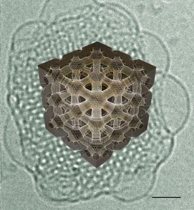
3-D reconstruction of one of the 2 water channels of an inverse bicontinuous cubic phase obtained from cryo-electron tomography experiments. Scale bar: 40 nm Credit: Davide Demurtas/EPFL
EPFL scientists, working with Nestlé, have now been able to study the 3D structure of cubosomes in detail for the first time. Published in Nature Communications, the breakthrough can help and promote the use of cubosomes in medicine and food science.
Molecules of a drug or a nutrient contained inside a cubosome can move by using the numerous tiny channels that make up its structure's interior.
The pharmaceutical industry already uses a similar system for drug delivery: the liposomes, which are also made of fats but in the shape of a sphere. Their intricate internal channels give cubosomes a very high internal surface, which offers great potential for the controlled delivery of nutrients and drugs.
In short, the properties of cubosomes, like other lipid-based delivery vessels, depend on their particular structures. The problem is that cubosomes are self-assembled, occurring 'spontaneously' after putting together the right ingredients (generally fats and a detergent) under the right conditions.
This means that scientists have limited control over their final structure, which makes it hard to optimize their design. In addition, it is very difficult to 'see' the interior of a cubosome and map out the various arrangements of its channels.
Davide Demurtas and Cécile Hébert from EPFL's Interdisciplinary Centre for Electron Microscopy (CIME), working with Laurent Sagalowicz at the Nestlé Research Center in Lausanne, have now uncovered the interior 3D structure of cubosomes, and have successfully matched their real-life findings to computer simulations.
The researchers used a microscopy technique called 'cryo-electron tomography' (CET). Their method involves embedding cubosomes in a type of 'glass' ice that does not form crystals, which would damage the cubosomes. The samples are kept at -170oC. The microscope then takes photographs while tilting the cubosome at different angles. The technique, which was carried out at CIME, can reconstruct the three-dimensional information to create images of the cubosomes in their native state and with unprecedented detail.
“This method allows us to get information about everything, both the inside and outside of the cubosomes,” says Cécile Hébert. “Because the CET microscope distinguishes the different densities between cubosome and ice, it is very sensitive and precise.”
The CET images clearly showed the internal cubic structure, as well as the internal 3D organization of the channels. The researchers also compared the images to the prevailing mathematical models used to make computer simulations of the interface between the interior and exterior. The real-life data successfully matched the theory.
“With this approach we can now forge a new understanding of the structure of the cubosomes' interior,” says Davide Demurtas. The success is expected to make the study and design of cubosomes with controlled macroscopic properties (e.g. controlled release) easier.
###
This work represents a collaboration of EPFL's Interdisciplinary Centre for Electron Microscopy (CIME) with the Nestlé Research Center Lausanne, EPFL's Institute of Cancer Research, and the Department of Health Science & Technology of ETH Zurich.
Reference
Demurtas D, Guichard P, Martiel I, Mezzenga R, Hébert C, Sagalowicz L. Direct visualization of dispersed lipid bicontinuous cubic phases by cryo-electron tomography. Nature Communications 17 Nov. 2015. DOI: 10.1038/NCOMMS9915.
Media Contact
All latest news from the category: Life Sciences and Chemistry
Articles and reports from the Life Sciences and chemistry area deal with applied and basic research into modern biology, chemistry and human medicine.
Valuable information can be found on a range of life sciences fields including bacteriology, biochemistry, bionics, bioinformatics, biophysics, biotechnology, genetics, geobotany, human biology, marine biology, microbiology, molecular biology, cellular biology, zoology, bioinorganic chemistry, microchemistry and environmental chemistry.


Newest articles

High-energy-density aqueous battery based on halogen multi-electron transfer
Traditional non-aqueous lithium-ion batteries have a high energy density, but their safety is compromised due to the flammable organic electrolytes they utilize. Aqueous batteries use water as the solvent for…

First-ever combined heart pump and pig kidney transplant
…gives new hope to patient with terminal illness. Surgeons at NYU Langone Health performed the first-ever combined mechanical heart pump and gene-edited pig kidney transplant surgery in a 54-year-old woman…

Biophysics: Testing how well biomarkers work
LMU researchers have developed a method to determine how reliably target proteins can be labeled using super-resolution fluorescence microscopy. Modern microscopy techniques make it possible to examine the inner workings…

