Highly automated live cell imaging speeds up the search for new drugs
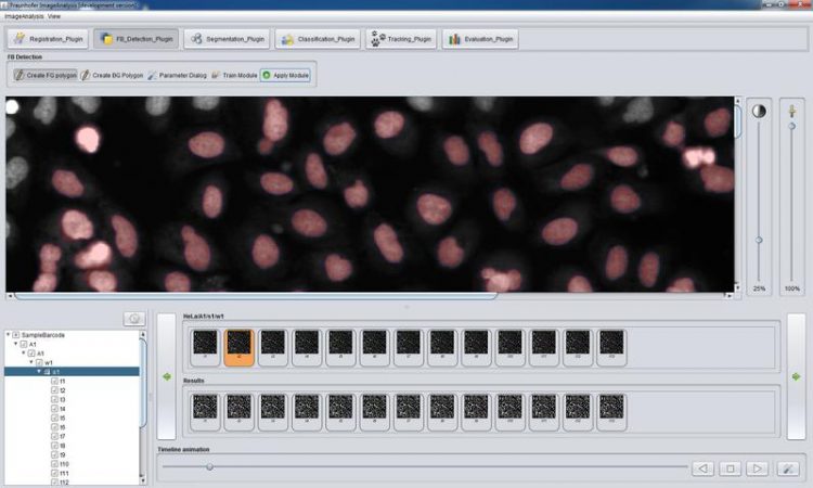
Zeta's clean graphical user interface. (c) Fraunhofer FIT
The new Zeta software platform allows researchers to implement specific imaging workflows for a broad range of applications in drug research very easily. The software was developed specifically for high content analysis of live cell imaging data that monitor and record the complete life cycle of cells.
The particular challenge for image analysis here is to detect the different phases of cell modification and cell division, and to record their temporal relationship. On this basis, a special visualization tool makes it easy to explore the data, to find individual differences and to determine the causes for different reactions of the cells.
“Using Zeta, researchers can analyze complex processes in the division of cells very easily and intuitively. A simple user interface guides them through the entire analysis workflow. And due to the evolution of Zeta into a modular software platform, we can now implement new applications much faster and thus at lower cost for our clients”, Dr. Andreas Pippow, Fraunhofer Institute for Applied Information Technology FIT, points out the main advantages of the latest Zeta version.
New software interfaces in the new version make it easier to integrate Zeta into all-encompassing high content analysis workflows. Imaging software often exists only as an isolated application – which was true also for earlier Zeta versions. What users need, however, is full integration with image data management and statistical analyses. Only if all steps in the entire workflow are supported by one coherent system, the users can freely explore and exploit their data. The latest Zeta version is a significant step in this direction.
At MipTec, September 23 to 25, 2014, the Zeta developers from Fraunhofer FIT demo a Zeta application that determines the cell division rate in cell assays. It is currently being used in cancer research in a large German pharmaceutical company.
The FIT researchers also demo a Zeta application in the study of physiological functions in tissue samples. While both applications are used in the quest for new pharmaceutical agents, they apply completely different image analysis methods. In the first application, fluorescent markers identify the cells; in the second, the objects must be detected without any specific marking.
Contact:
Alex Deeg
pr@fit.fraunhofer.de
Phone +49 2241 14-2208
Media Contact
More Information:
http://www.fit.fraunhofer.deAll latest news from the category: Information Technology
Here you can find a summary of innovations in the fields of information and data processing and up-to-date developments on IT equipment and hardware.
This area covers topics such as IT services, IT architectures, IT management and telecommunications.


Newest articles

Properties of new materials for microchips
… can now be measured well. Reseachers of Delft University of Technology demonstrated measuring performance properties of ultrathin silicon membranes. Making ever smaller and more powerful chips requires new ultrathin…

Floating solar’s potential
… to support sustainable development by addressing climate, water, and energy goals holistically. A new study published this week in Nature Energy raises the potential for floating solar photovoltaics (FPV)…

Skyrmions move at record speeds
… a step towards the computing of the future. An international research team led by scientists from the CNRS1 has discovered that the magnetic nanobubbles2 known as skyrmions can be…

