TB bacteria mask their identity to intrude into deeper regions of lungs
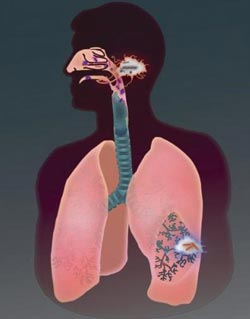
Flying under the radar: tuberculosis-causing mycobacteria initiate infection in the lower lung to evade pathogen-killing cells.<br><br>Credit: Ramakrishnan Lab/University of Washington<br>
TB-causing bacteria appear to mask their identity to avoid recognition by infection-killing cells in the upper airways. The bacteria call up more permissive white blood cells in the deeper regions of the lungs and hitch a ride inside them to get into the host's body.
Details on this finding are reported Dec. 16 in the advanced online edition of the journal Nature. The research was a collaboration between the University of Washington and the Seattle Biomedical Research Institute.
Dr. Lalita Ramakrishan, who studies how TB evades the body's immune system and manipulates the body's defenses for its own ends, is the senior author. She is a UW professor of microbiology, medicine and immunology. The lead author is C.J. Cambier of the UW Department of Immunology.
Ramakrishnan noted that the recent study also suggests an explanation for the longstanding observation that tuberculosis infections begin in the comparatively sterile lower lungs. In the upper respiratory tract, resident microbes and inhaled microbes of a variety of species signal their presence.
These tip-offs alert and attract many infection-fighting cells to the upper airways. The presence of other microbes in the upper airway may thereby help to keep TB infections at bay by creating a hostile environment.
Their presence may explain why TB is a less contagious disease than those caused by several other respiratory pathogens.
To cause disease, TB bacteria must sneak through this well-patrolled area and head for parts of the lungs where fewer microbiocidal cells are policing.
Almost like intruders wearing a stocking over their faces to keep surveillance cameras from clearly recording their features, the TB pathogens produce particular types of fatty substances, or lipids, on their cell surfaces.
These lipids, abbreviated as PDIM, are already known to be associated with bacterial virulence. The researchers showed that PDIM lipids function by masking the underlying molecular patterns that would reveal their dangerous nature to macrophages, a first-line defense of infection-fighting cells.
At the same time, a related lipid – called PGL – on the bacterium's cell surface promotes the recruitment of cells described as permissive macrophages. These clean-up cells engulf but don't kill the TB pathogens. Instead, they take them across the lung lining, deep into the lung tissue where the bacteria can establish an infection.
According to the researchers on this study, these mechanisms appear to allow certain TB pathogens to avoid detection by the pattern recognition receptors that enable some infection-fighting cells to spot a variety of different disease microbes through the pathogen-associated molecular patterns on or near their cell surface.
Like most other bacteria, TB pathogens have many of these telltale molecular patterns that should activate an immune response. However, TB pathogens have evolved mechanisms to circumvent tripping the alarm, in this case by physically masking the otherwise detectable pattern. This cover-up allows them to infect the airway initially by avoiding the infection-fighting cell populations that are detrimental to their survival, the researchers noted.
The TB pathogens then use the other lipid molecule, PGL, to co-opt a host chemical pathway that triggers the recruitment of the permissive macrophages.
The present study expands on earlier work in the Ramakrishan and collaborative labs, which helped describe the strategies by which TB pathogens manipulate host pathways for their own purposes after they enter certain host cells.
These include the secretion of proteins that help expand the niche for TB by recruiting macrophages to the early lung tubercles characteristic of the disease. The present study describes earlier stages in infection, when the pathogens first come in contact with their potential host at the surface of the lining of the lung.
“The current study suggests the manner in which the TB pathogens manipulate recruitment of the first responding macrophages to gain access to their preferred niche,” the researchers noted.
“The choreographed entry involves two related TB cell lipids acting in concert to avoid one host pathway while inducing another,” they wrote. The findings link the previously known, absolutely essential virulence factor on the surface of TB cells, PDIM, to the evasion of immune cell detection. They also might explain why a certain pathogen molecular pattern recognition system is dispensable in protecting against TB. On the other hand, PGL is not required on the surface of TB cells for them to infect the body.
Ramakrishnan noted that globally, a lot of samples of TB taken from infected patients do not have PGL. “However,” she and her research team noted, “the importance of PGL in mediating TB virulence or transmission is underscored by its presence in many of the W-Beijing strains” of TB which are starting to rapidly appear in more patient samples, and which have predominated in outbreaks in North America.
Ramakrishnan explains that their findings suggest how PGL may play an important role in increasing TB's infectivity.
“The presence of PGL in ancestral strains of TB suggest it played an integral role in the evolution of TB infectivity,” the researchers noted. “TB is an ancient disease and the enhanced infectivity conferred by PGL may have been essential for most of its history before human crowding, with its increased opportunity for transmission, made it dispensable.”
The study findings, and previous work on TB, might also explain why smaller droplets of TB are more infectious than larger ones. Only the smaller droplets can make their way down into the lower airways. On the flip side, all it takes is 3 or fewer TB mycobacteria with PGL-producing ability to enter the lower lungs and start an infection.
The other researchers on the study, in addition to Cambier and Ramkrishnan, were Kevin K. Takaki, David M. Tobin, and Christina L. Cosma, all of the UW Department of Microbiology; Ryan Larson and Kevin N. Urdahl of the of the UW Department of Immunology and the Seattle Biomedical Research Institute. Urdahl also is from the UW Department of Pediatrics.
The research was supported by training and research grants from the National Science Foundation, American Lung Association, National Institutes of Health, and American Cancer Society. Tobin is an NIH New Innovator and Ramakrishnan is an NIH Pioneer.
Media Contact
More Information:
http://www.uw.eduAll latest news from the category: Health and Medicine
This subject area encompasses research and studies in the field of human medicine.
Among the wide-ranging list of topics covered here are anesthesiology, anatomy, surgery, human genetics, hygiene and environmental medicine, internal medicine, neurology, pharmacology, physiology, urology and dental medicine.


Newest articles
Machine learning algorithm reveals long-theorized glass phase in crystal
Scientists have found evidence of an elusive, glassy phase of matter that emerges when a crystal’s perfect internal pattern is disrupted. X-ray technology and machine learning converge to shed light…
Mapping plant functional diversity from space
HKU ecologists revolutionize ecosystem monitoring with novel field-satellite integration. An international team of researchers, led by Professor Jin WU from the School of Biological Sciences at The University of Hong…
Inverters with constant full load capability
…enable an increase in the performance of electric drives. Overheating components significantly limit the performance of drivetrains in electric vehicles. Inverters in particular are subject to a high thermal load,…

