Retinoic acid gradient visualized for the first time in an embryo
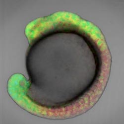
These are retinoic acid gradients in a zebrafish embryo, visualized using cyan and yellow fluorophores. Credit: RIKEN<br>
This technique enabled them to observe two concentration gradients going in opposing directions along the head-to-tail axis of the embryo, thus providing long-awaited evidence that retinoic acid is a morphogen.
The report, published today in the journal Nature, puts an end to a long-standing debate around the presence of retinoic acid gradients across the vertebrate embryo, during the early stages of development. It also sheds light on the role of retinoic acid in tissue development.
Retinoic acid has been thought to be a morphogen, a signalling molecule that diffuses throughout the embryo switching genes on and off and imparting different cell fates depending on its concentration. However, retinoic acid concentration gradients had never been visualized because retinoic acid cannot be tagged with the commonly used 'green fluorescent protein' GFP, or GFP-like proteins, as label.
“Until now no one had succeeded in monitoring the concentration of retinoic acid in real-time in a live embryo, and there was no direct data proving the existence of a retinoic acid gradient in the vertebrate embryo, explains Dr. Miyawaki, who led the research.
In order to monitor the concentration of retinoic acid in live zebrafish embryos at the early stages of their development, Dr. Miyawaki and his colleague Dr. Shimozono developed a technique to tag the molecule that acts as receptor for retinoic acid with genetically-encoded, coloured fluorophores. Based on the principle of fluorescence resonance energy transfer (FRET), the tags allow them to visualize the presence of retinoic acid and quantitatively determine its concentration over time.
By combining this technique with pharmacological and genetic manipulations, Miyawaki and his team demonstrate the presence of two linear retinoic acid concentration gradients across the antero-posterior axis of the embryo, from the trunk area to the head and the tail. Their findings suggest that retinoic acid diffuses quickly, thus establishing stable and robust gradients that are resistant to external perturbations.
“A better understanding of the gradients of retinoic acid is essential for research into the patterns of tissue development. It is necessary if we ever want to control the development of three-dimensional tissue structures from induced pluripotent stem cells, for regenerative medicine for example,” concludes Dr. Miyawaki.
Media Contact
More Information:
http://www.riken.jpAll latest news from the category: Life Sciences and Chemistry
Articles and reports from the Life Sciences and chemistry area deal with applied and basic research into modern biology, chemistry and human medicine.
Valuable information can be found on a range of life sciences fields including bacteriology, biochemistry, bionics, bioinformatics, biophysics, biotechnology, genetics, geobotany, human biology, marine biology, microbiology, molecular biology, cellular biology, zoology, bioinorganic chemistry, microchemistry and environmental chemistry.


Newest articles

“Nanostitches” enable lighter and tougher composite materials
In research that may lead to next-generation airplanes and spacecraft, MIT engineers used carbon nanotubes to prevent cracking in multilayered composites. To save on fuel and reduce aircraft emissions, engineers…

Trash to treasure
Researchers turn metal waste into catalyst for hydrogen. Scientists have found a way to transform metal waste into a highly efficient catalyst to make hydrogen from water, a discovery that…

Real-time detection of infectious disease viruses
… by searching for molecular fingerprinting. A research team consisting of Professor Kyoung-Duck Park and Taeyoung Moon and Huitae Joo, PhD candidates, from the Department of Physics at Pohang University…

