Molecular 3D-maps unlock new ways of studying human reproduction
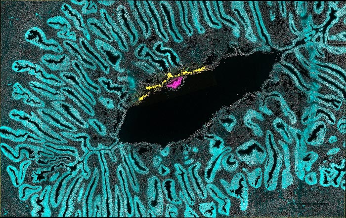
The embryonic disc - which will give rise to the fetus, and the emerging amniotic sac are in pink. The early placenta, acting as an intermediary between embryo and mother, is in yellow. The uterus is in cyan.
Credit: University of Cambridge
Scientists have identified the biochemical signals that control the emergence of the body pattern in the primate embryo. This will guide work to understand birth defects and pregnancy loss in humans.
The study also provides a crucial reference for foetal tissue generation in the lab – such tissue is in short supply but is needed for drug screening and studies into stem cell-based treatments to regenerate body tissues in diseases like Parkinson’s, for example.
Embryos develop from a clump of cells into highly organised structures. However, until now the signals orchestrating this transformation have remained hidden from observation inside the womb.
Measuring gene activity in three dimensions, researchers have generated molecular maps of the second week of gestation as it has never been seen before. Their work is published today in the journal Nature.
“This work will provide a definitive laboratory reference for future studies of early embryo development, and the embryonic origins of disease,” said Dr Thorsten Boroviak in the University of Cambridge’s Department of Physiology, Development and Neuroscience and senior author of the study.
The second week of gestation is one of the most mysterious, yet critical, stages of embryo development. Failure of development during this time is one of the major causes of early pregnancy loss and birth defects.
In previous work, Boroviak showed that the first week of development in marmoset monkeys is remarkably similar to that in humans. But with existing methods he could not explore week two of development, after the embryo implants into the womb.
A new laser-assisted technique enabled the team to track down the earliest signals driving the establishment of the body axis – when the symmetrical structure of the embryo starts to change. One end becomes committed to developing into the head, and the other end becomes the ‘tail’.
The team discovered that asymmetric signals come from the embryo itself and from transient structures that support the embryo during its development – the amnion, yolk sac, and precursors of the placenta.
“Our virtual reconstructions show the developing embryo and its’ supporting tissues in the days after implantation in incredible detail,” said Boroviak.
The blueprint unlocks new ways of studying human reproduction and development. In the future, the team plans to use their new technique to investigate origins of pregnancy complications and birth defects using engineered embryo models. Understanding more about human development will help scientists to understand how it can go wrong and take steps towards being able to fix problems.
The pre-implantation period, before the developing embryo implants into the mother’s womb, has been studied extensively in human embryos in the lab. On the seventh day the embryo implants into the womb to survive and develop. Very little was previously known about the development of the human embryo once it implants, because it becomes inaccessible for study.
Boroviak’s team used implanted embryos of the marmoset, a small New World monkey, in their study because they are very similar to human embryos at this early stage of development.
Journal: Nature
DOI: 10.1038/s41586-022-04953-1
Subject of Research: Animals
Article Title: Spatial profiling of early primate gastrulation in utero
Article Publication Date: 16-Jun-2022
All latest news from the category: Life Sciences and Chemistry
Articles and reports from the Life Sciences and chemistry area deal with applied and basic research into modern biology, chemistry and human medicine.
Valuable information can be found on a range of life sciences fields including bacteriology, biochemistry, bionics, bioinformatics, biophysics, biotechnology, genetics, geobotany, human biology, marine biology, microbiology, molecular biology, cellular biology, zoology, bioinorganic chemistry, microchemistry and environmental chemistry.


Newest articles

“Nanostitches” enable lighter and tougher composite materials
In research that may lead to next-generation airplanes and spacecraft, MIT engineers used carbon nanotubes to prevent cracking in multilayered composites. To save on fuel and reduce aircraft emissions, engineers…

Trash to treasure
Researchers turn metal waste into catalyst for hydrogen. Scientists have found a way to transform metal waste into a highly efficient catalyst to make hydrogen from water, a discovery that…

Real-time detection of infectious disease viruses
… by searching for molecular fingerprinting. A research team consisting of Professor Kyoung-Duck Park and Taeyoung Moon and Huitae Joo, PhD candidates, from the Department of Physics at Pohang University…

