Cartography of the visual cortex: Charting a new course for the organization of visual space
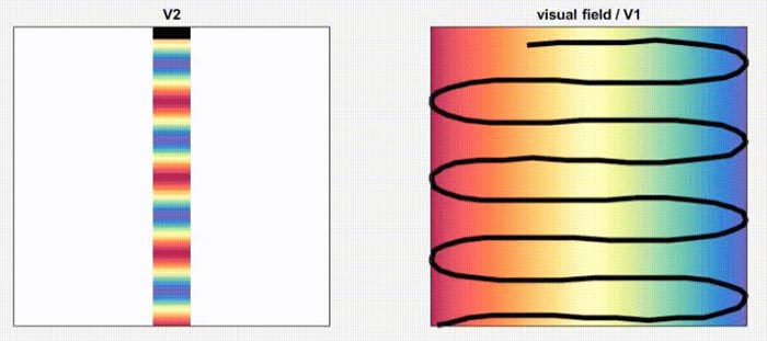
The featured video shows the sinusoidal transformation between the visual field and V2. It shows how regions in the visual field are mapped onto regions in V2. The colors correspond to the azimuth (left/right) axis of the visual field.
Credit: Max Planck Florida Institute for Neuroscience
Researchers at the Max Planck Florida Institute for Neuroscience (MPFI) have uncovered a surprisingly complex yet precisely ordered map of visual space in area V2 of the cortex.
Requiring a discerning eye, mathematical precision, and keen sense of aesthetics, map making is a unique application of both art and science. Though the scale may differ, neuroscientists that study vision are like cartographers of the brain; investigating and mapping how our brain represents and makes sense of what we see in the world. The visual cortex, a specialized region responsible for visual processing, contains intricate neural circuits that evaluate information arriving from our eyes and preferentially respond to distinguishing visual features such as color, edges, motion, and location in visual space. Despite the sheer complexity of this information, our brains do a remarkable job of efficiently organizing neurons together, helping us to better understand our visual landscapes.
One major organizational property the visual cortex employs is called retinotopic mapping, where neurons within are arranged in an orderly way that preserves the spatial information arriving from the retina (light sensing portion of the eye). Much like the Mercator projection is to cartography, retinotopic maps in the visual cortex are thought to follow a widely adopted and well-characterized pattern. The prevailing theory is that brain areas like the primary visual cortex (V1) follow a smooth and simple method of mapping. What you see is what you get; objects in visual space that activate portions of the retina, will light up neurons in an identical pattern in the brain.
Despite the wealth of evidence in multiple species supporting this type of linear mapping, small hints and discrepancies existed within previous studies that suggested the possibility of other arrangements. The question remained, do additional methods of spatial mapping exist in the brain?
Shedding light on this question and challenging the prevailing theory, researchers in MPFI’s Fitzpatrick Lab have uncovered for the very first time a new type of spatial mapping within the secondary area (V2) of the visual cortex. Published recently in Neuron, the team employed a combination of single-cell functional imaging, computational modeling and connectivity studies, to reveal a sinusoidal or wavelike organization in area V2 of the tree shrew. Their surprising insight has deepened our understanding of neural representations of visual space and underlined the importance of precise retinotopic mapping in the visual cortex.
Madineh Sedigh-Sarvestani, Ph.D., a postdoctoral researcher at MPFI and first author of the publication, joined the Fitzpatrick lab interested in understanding the organization, function, and behavioral link of visual areas beyond the well-studied V1. Her investigation began in V1’s closely related neighbor, V2, a visual area that has been extensively studied in primates but less so in animals amenable to recent genetic tools developed in mice. The tree shrew perfectly fits this criterion, as it’s a close relative of primates and has a smooth brain ideal for imaging. Utilizing high resolution calcium imaging, Sedigh-Sarvestani expected to find a map of visual space very similar to V1’s golden-standard.
Presenting tree shrews with visual stimuli that varied in position within the visual field, the team mapped the corresponding neurons in V2 that lit up in response to a visual stimulus’ location in space. What they discovered was two very distinct maps in V2. The map of an object’s elevation, how high or low it is, followed closely with the smooth linear map found in V1 but mapping the azimuth, its horizontal position left or right of center, revealed a dramatically different sinusoidal, or oscillating pattern. But why would simple spatial maps exist in V1 and more complex in V2, could differences in the regions’ shapes play a role? To answer this question the MPFI researchers turned to computer modeling to recreate the conditions found within the brain, with the goal of producing a spatial map that optimizes coverage of the visual field. By varying only shape, the algorithm found that the optimal spatial map for the square V1 region followed the smooth, linear arrangement but for the thin, elongated V2, a sinusoidal map emerged corroborating previous results. Cementing this idea, the MPFI team, led by the lab’s histology coordinator Nicole Shultz, used colored dyes to trace the connections from V1 to the V2 region, finding that the neuronal projections from V1 perfectly aligned with the sinusoidal map in V2.
“Our results demonstrate that orderly organization of visual space in the brain, does not necessarily have to follow the guiding principles we are accustomed to thinking about,” notes David Fitzpatrick, Ph.D., CEO and Scientific Director of MPFI. “Though this organization may be less straightforward than we originally thought, it still has remarkable and beautiful order.”
Beyond this intriguing finding, researchers in the Fitzpatrick lab made one more critically important discovery with broad implications for the field of visual neuroscience; neuronal preference for certain visual features is tied directly to the retinotopic map of visual space. Predominantly thought to be independent organizational principles, the MPFI team demonstrated their interconnectedness by studying the response properties of neurons in V2 for binocular or monocular stimuli. They found that the oscillating map of visual space completely overlapped with the functional feature map, illustrating that the sensitivity neurons have for visual features is not uniform but can vary depending on where the features are in visual space.
“This type of synergy between these two principles, preference for visual features and their location in space, starts to reveal unique information about the behavior or environment of certain animals,” describes Sedigh-Sarvestani. “The wiring of visual circuits that determine the patterns that end up in the brain are influenced by our visual experience; What you see and where you see it.”
In the future, Sedigh-Sarvestani plans to investigate if other visual features are tethered to mapping of visual space in different regions of the visual cortex and if this organization can be traced back to the retina and eventually, an animal’s native environment and their movements within that environment; leading to a more comprehensive understanding of visual perception.
“What we found really forced us to rethink how maps of visual space are formed and to recognize that neural circuits in the visual cortex can be functionally specialized for different regions of visual space,” explains Fitzpatrick. “Our findings open the door to a different way of thinking about how cortical circuits are organized, how they contribute to visual perception, and ultimately, behavior.”
This work was supported by the National Institutes of Health Grants, and the Max Planck Florida Institute for Neuroscience. The content of this article is solely the responsibility of the authors and does not necessarily represent the official views of the funding agencies.
Journal: Neuron
DOI: 10.1016/j.neuron.2021.09.053
Method of Research: Experimental study
Subject of Research: Animals
Article Title: Sinusoidal transformation of the visual field is the basis for periodic maps in area V2
Article Publication Date: 22-Oct-2021
Media Contact
Helena Decker
Max Planck Florida Institute for Neuroscience
helena.decker@mpfi.org
Cell: 561-3019661
All latest news from the category: Life Sciences and Chemistry
Articles and reports from the Life Sciences and chemistry area deal with applied and basic research into modern biology, chemistry and human medicine.
Valuable information can be found on a range of life sciences fields including bacteriology, biochemistry, bionics, bioinformatics, biophysics, biotechnology, genetics, geobotany, human biology, marine biology, microbiology, molecular biology, cellular biology, zoology, bioinorganic chemistry, microchemistry and environmental chemistry.


Newest articles
Combatting disruptive ‘noise’ in quantum communication
In a significant milestone for quantum communication technology, an experiment has demonstrated how networks can be leveraged to combat disruptive ‘noise’ in quantum communications. The international effort led by researchers…

Stretchable quantum dot display
Intrinsically stretchable quantum dot-based light-emitting diodes achieved record-breaking performance. A team of South Korean scientists led by Professor KIM Dae-Hyeong of the Center for Nanoparticle Research within the Institute for…

Internet can achieve quantum speed with light saved as sound
Researchers at the University of Copenhagen’s Niels Bohr Institute have developed a new way to create quantum memory: A small drum can store data sent with light in its sonic…

